Research Article · Volume 14, Issue 11, 30 September 2026 · Open Access
Phylogenetic Analysis of Feline Coronavirus (FCoV) Isolated from Tangerang, Indonesia Based on the Nucleocapsid (N) and Membrane (M) Genes
· · · · · · ·
Affiliations & Notes ⌄
1Study Program of Veterinary Medicine, School of Veterinary Medicine and Biomedical Science, IPB University, Bogor, Indonesia
2Division of Physiology, School of Veterinary Medicine and Biomedical Science, IPB University, Bogor, Indonesia
3Division of Anatomy, Histology, and Embriology, School of Veterinary Medicine and Biomedical Science, IPB University, Bogor, Indonesia
4Study Program of Animal Biomedical Science, Postgraduate School, IPB University, Bogor, Indonesia
5Division of Medical Microbiology, School of Veterinary Medicine and Biomedical Science, IPB University, Bogor, Indonesia. *Correspondence | Okti Nadia Poetri, Division of Medical Microbiology, School of Veterinary Medicine and Biomedical Science, IPB University, Bogor, Indonesia
The identity of the amplified products as FCoV N and M gene sequences was further supported by sequencing and BLASTn analysis, which demonstrated moderate nucleotide identity with FCoV reference sequences (Table 2).
BLASTn analysis showed that the N gene shared 91.22–94.23% nucleotide identity with the Chinese genotype I reference strain PV246293, whereas the M gene exhibited 92.63–94.43% identity with the Chinese genotype I reference strain PP444763.
This study characterized partial membrane (M) and nucleocapsid (N) gene sequences of FCoV from Tangerang, Indonesia.
BLASTn and phylogenetic analyses showed genetic relatedness between the Tangerang sequences and genotype I reference strains from different geographical regions.
Selected from the article's own text.
Abstract
Feline coronavirus (FCoV) is a positive-sense single-stranded RNA virus that is widely distributed in the cat population of Indonesia. FCoV infection typically produces subclinical or mild gastrointestinal signs; however, high mutation rates facilitate systemic dissemination and unpredictable disease progression to feline infectious peritonitis (FIP), which posing a significant threat to feline health.This study aimed to perform molecular characterization and phylogenetic analysis of FCoV detected in symptomatic cats in Tangerang, Indonesia, based on two structural genes, the membrane (M) and nucleocapsid (N) genes. Eleven archived samples previously identified as FCoV-positive based on the spike (S) gene were analyzed using reverse transcription-polymerase chain reaction (RT-PCR) followed by Sanger sequencing and phylogenetic mapping. Of the 11 archived samples previously identified as FCoV-positive, seven (63.6%) were successfully amplified and sequenced for both the N and M genes. BLASTn analysis showed that the N gene shared 91.22–94.23% nucleotide identity with the Chinese genotype I reference strain PV246293, whereas the M gene exhibited 92.63–94.43% identity with the Chinese genotype I reference strain PP444763. Phylogenetic analysis placed all Tangerang sequences within genotype I. The N gene sequences formed a distinct monophyletic clade, whereas the M gene sequences were resolved into two closely related lineages. These findings provide baseline molecular epidemiological data on FCoV circulating in Tangerang and may support future surveillance and studies of its genetic diversity and evolution in Indonesia.
Feline coronavirus (FCoV) is an enveloped, positive-sense RNA virus in the family Coronaviridae and genus Alphacoronavirus that infects cats worldwide. Pathogenetically, FCoV has traditionally been classified into two biotypes: Feline enteric coronavirus (FECV), which is generally associated with asymptomatic infection or mild enteric disease, and Feline infectious peritonitis virus (FIPV), a highly pathogenic biotype responsible for an often fatal systemic disease ; . Beyond pathogenicity, FCoV is genetically distinguished into genotype I, the most dominant type in the field, and genotype II, which is a result of recombination between genotype I FCoV and Canine coronavirus (CCoV) .
FCoV infection exhibits highly efficient transmission and high global prevalence, reaching 47.6% in Europe and up to 67.9% in Asian regions such as Fujian, China ; . The evolutionary dynamics of this virus are heavily influenced by genetic recombination and in vivo mutations, such as the emergence of FIPV from FECV. Recent epidemiological events, such as the 2023 outbreak in Cyprus involving the recombinant FCoV-23 variant, demonstrated increased transmission efficiency and neurological manifestations . These developments underscore the role of various animal species as reservoirs and highlight the necessity of molecular studies to understand viral adaptation ; .
In Indonesia, studies regarding the prevalence and characterization of FCoV variants remain very limited. Previous reports indicate FCoV antibody prevalence as high as 90% in 10 cats in Yogyakarta, along with molecular detection in imported cats ; . Despite this high prevalence, updated information on the genetic variation of local Indonesian strains is scarce. Understanding the molecular structure of the FCoV genome comprising the spike (S), envelope (E), membrane (M), and nucleocapsid (N) genes is essential for epidemiological studies .
The limited molecular data available for Indonesian FCoV strains, particularly for the M and N genes, restrict our understanding of their genetic diversity and molecular epidemiology. The M protein is an integral membrane protein that plays an important role in coronavirus assembly, budding, and virion morphogenesis through its interactions with other structural proteins. Meanwhile, the N protein binds to the viral genomic RNA to form the ribonucleoprotein complex and contributes to viral RNA replication, transcription, and virion assembly . Despite their important biological roles, molecular information on the M and N genes of Indonesian FCoV strains remains limited, particularly for viruses circulating in the Tangerang region. Therefore, analysis of partial sequences of the M and N genes may provide additional information on the genetic characteristics and phylogenetic relationships of local FCoV strains.
To address these knowledge gaps, this study aimed to detect FCoV in clinical samples collected from cats in Tangerang, Indonesia, and to characterize the partial sequences of the membrane (M) and nucleocapsid (N) genes of the detected viruses. The specific objectives were to evaluate sequence variation among the Indonesian strains and determine their phylogenetic relationships with representative FCoV sequences available in public databases. The findings provide baseline molecular epidemiological data on FCoV circulating in Tangerang and may support future surveillance, research on its genetic diversity and evolution in Indonesia, and the selection of locally relevant genomic regions for further diagnostic investigation.
MATERIALS AND METHODS
Archived samples
A total of 11 archived samples, including feces, rectal swabs, and ascites fluid, were utilized in this study. The samples were originally collected from cats showing clinical signs suggestive of feline infectious peritonitis (FIP) and had been included in our previous study investigating FCoV based on partial spike (S) gene sequences . The present study represents a distinct molecular analysis of these archived samples, focusing on the partial nucleocapsid (N) and membrane (M) gene sequences. The archived samples were maintained at the Immunology Laboratory, Division of Medical Microbiology, School of Veterinary Medicine and Biomedical Sciences, IPB University, and stored frozen at −20 °C until further molecular analysis.
Viral rna extraction
Total viral RNA was isolated using the Geneaid Viral Nucleic Acid Extraction Kit II (New Taipei City, Taiwan) according to the manufacturer’s protocol. Briefly, 200 μL of sample was lysed using 400 μL of VB Lysis Buffer and incubated for 10 minutes at room temperature. Nucleic acid binding was performed by adding 450 μL of AD Buffer (pre-mixed with absolute ethanol) and passing the mixture through a VB Column via centrifugation at 18,626 × g. The column was subsequently washed with W1 and Wash buffers to remove contaminants. Finally, the RNA was eluted into 50 μL of RNase-free water and stored at -20 °C until further use. Viral RNA concentration was not measured quantitatively in this study.
Synthesis of CDNA
The extracted RNA was converted into complementary DNA (cDNA) using the ReverTra Ace-α-™ kit (TOYOBO, Osaka, Japan). The cDNA synthesis for the partial N and M gene sequences was performed separately using their respective gene-specific reverse primers. For the nucleocapsid (N) gene, a specific reverse primer FCoV-N-R (5′-TGCGTTTAGTTCGTAACCTC-3′) was used, while for the membrane (M) gene, the primer FCoV-M-R (5′-GGAAGGTTCATCTCCCCAGTTGACG-3′) was employed. The reaction mixture consisted of 5x RT buffer, a dNTP mixture, an RNase inhibitor, a specific primer, and 1 μL of ReverTra Ace enzyme. Synthesis was performed at 42 °C for 20 minutes, followed by enzyme inactivation at 99 °C for 5 minutes. Viral cDNA concentration was not measured quantitatively in this study.
Polymerase chain reaction (PCR) amplification
Amplification was conducted using KOD One™ PCR Master Mix -BLUE- (TOYOBO, Osaka, Japan) at a 25 μL reaction volume. A previously confirmed FCoV-positive sample from our earlier study was included as the positive control, while a no-template control (NTC), in which the template was replaced with nuclease-free water, was included as the negative control in each PCR run. Specific primers were used to amplify partial sequences of the N and M genes, with expected amplicon sizes of 1202 bp and 880 bp, respectively, as shown in Table 1. The thermal cycling conditions for the N gene included initial denaturation at 95 °C for 3 minutes, followed by 35 cycles of denaturation (94 °C, 30s), annealing (56 °C, 30s), and extension (72 °C, 75s). For the M gene, the annealing temperature was adjusted to 58 °C with an extension time of 60 seconds. A final extension was performed at 72 °C for 10 minutes.
Electrophoresis and sequencing
PCR products were analyzed by 1.5% agarose gel electrophoresis (modified from at 80–150 V for 35 minutes. Gels were stained with ethidium bromide and visualized using a UV transilluminator. PCR products showing bands corresponding to the expected amplicon sizes for the partial N and M gene sequences were selected for bidirectional Sanger sequencing at Apical Scientific Sdn. Bhd. (1st BASE), Malaysia, for bidirectional Sanger sequencing. The identity of the amplified products was subsequently verified through sequence analysis.
Phylogenetic and data analysis
Sequencing data were analyzed using MEGA (Molecular Evolutionary Genetics Analysis) version 12.1.1 . Chromatograms were visually inspected for quality, and forward and reverse sequences were assembled into a consensus sequence after trimming primer-binding regions. Multiple sequence alignment was performed using ClustalW alongside global reference sequences from GenBank. Two separate phylogenetic trees, one based on partial M gene sequences and the other on partial N gene sequences, were constructed using the neighbor-joining method and the Kimura 2-parameter model with 1,000 bootstrap replicates. The resulting trees were visualized using iTOL (Interactive Tree of Life) . BLASTn (Basic Local Alignment Search Tool Nucleotide) analysis was used to assess nucleotide sequence similarity between the Indonesian strains and FCoV sequences available in GenBank. All samples had been previously classified as genotype I based on S gene analysis . The N and M gene phylogenies were used to assess broader genetic relationships, not for genotype assignment.
RESULTS
RT-PCR amplification of N and M genes
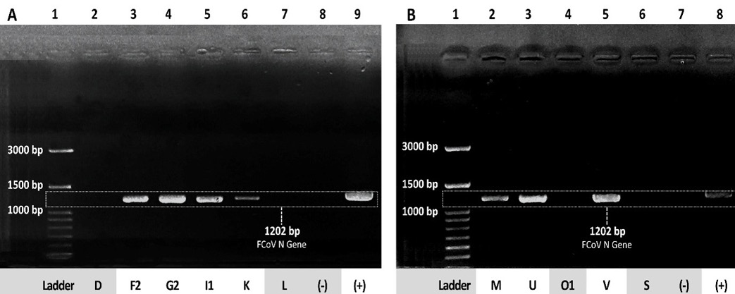
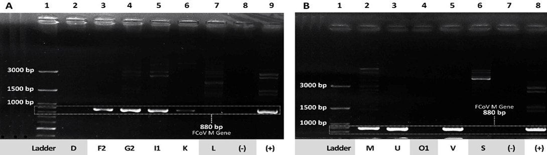
Molecular detection of Feline coronavirus (FCoV) was conducted on 11 clinical samples using RT-PCR targeting both the nucleocapsid (N) and membrane (M) genes. The results revealed that seven out of eleven samples (63.6%) were positive for both targets. Electrophoresis on a 1.5% agarose gel showed distinct bands at 1202 bp for the N gene (Figure 1) and 880 bp for the M gene (Figure 2).
The positive samples code were identified as F2 , G2 , I1, K , M, U, and V. Conversely, four samples (D, L, O1, and S) tested negative. Detailed sample information and RT-PCR results are summarized in Table 2.
Sequence alignment, variation, and BLASTn results
Bidirectional sequencing of the N gene amplicons yielded consensus sequences with lengths ranging from 788 to 797 bp after quality trimming and assembly of the forward and reverse reads. The final sequence lengths were 797 bp for F2, 794 bp for G2, 788 bp for I1, 796 bp for K, 794 bp for M, 789 bp for U, and 797 bp for V. For the M gene, analysis and consensus alignment of the chromatograms yielded final sequence lengths ranging from 768 to 771 bp whereas 771 bp for F2, G2, K, M, and V; 768 bp for I1 and U. BLASTn analysis against FCoV reference sequences available in GenBank showed nucleotide sequence identity values ranging from 91.22% to 94.23% for the N gene sequences, and between 92.63% and 94.43% for the M gene sequences (Table 2).
Table 1: Specific primers for FCoV N and M gene amplification.
Target Gene
Primer Name
Sequence (5′→3′)*
Fragment Size
Reference
N Gene
FCoV-N-F
GCACGTACTGAYAATTTGAGTGAAC
1202 bp
Shi et al., 2024
FCoV-N-R
TGCGTTTAGTTCGTAACCTC
M Gene
FCoV-M-F
CCCGACGAAGCATTYTTGTTTGAACTA
880 bp
Shi et al., 2024
FCoV-M-R
GGAAGGTTCATCTCCCCAGTTGACG
Footnotes:*Y = C or T according to the IUPAC nucleotide code.
Table 2: Summary of RT-PCR detection and molecular characteristics of partial N and M gene sequences from archived samples.
No.
Sample code
Sample type
Sequence name
N gene PCR
N sequence length (bp)
N nucleotide identityᵃ (%)
M gene PCR
M sequence length (bp)
M nucleotide identityᵇ (%)
1
D
Feces
NA
−
NA
NA
−
NA
NA
2
F2
Feces
FCOV/ID-TGR/F2/2024
+
797
94.23
+
771
94.43%
3
G2
Feces
FCOV/ID-TGR/G2/2024
+
794
93.22
+
771
93.65%
4
I1
Rectal swab
FCOV/ID-TGR/I1/2024
+
788
91.83
+
768
92.63%
5
K
Rectal swab
FCOV/ID-TGR/K/2024
+
796
94.10
+
771
94.43%
6
L
Rectal swab
NA
−
NA
NA
−
NA
NA
7
M
Rectal swab
FCOV/ID-TGR/M/2024
+
794
93.35
+
771
93.65%
8
U
Feces
FCOV/ID-TGR/U/2024
+
789
91.22
+
768
92.88%
9
O1
Rectal swab
NA
−
NA
NA
−
NA
NA
10
V
Feces
FCOV/ID-TGR/V/2024
+
797
93.32
+
771
94.43%
11
S
Ascitic fluid
NA
−
NA
NA
−
NA
NA
Footnotes: ᵃ Nucleotide sequence identity determined by BLASTn comparison with the FCoV sequence available in GenBank under accession number PV246293; ᵇ Nucleotide sequence identity determined by BLASTn comparison with the FCoV sequence available in GenBank under accession number PP444763; NA: Not applicable because no PCR product of the expected size was obtained.
Phylogenetic analysis
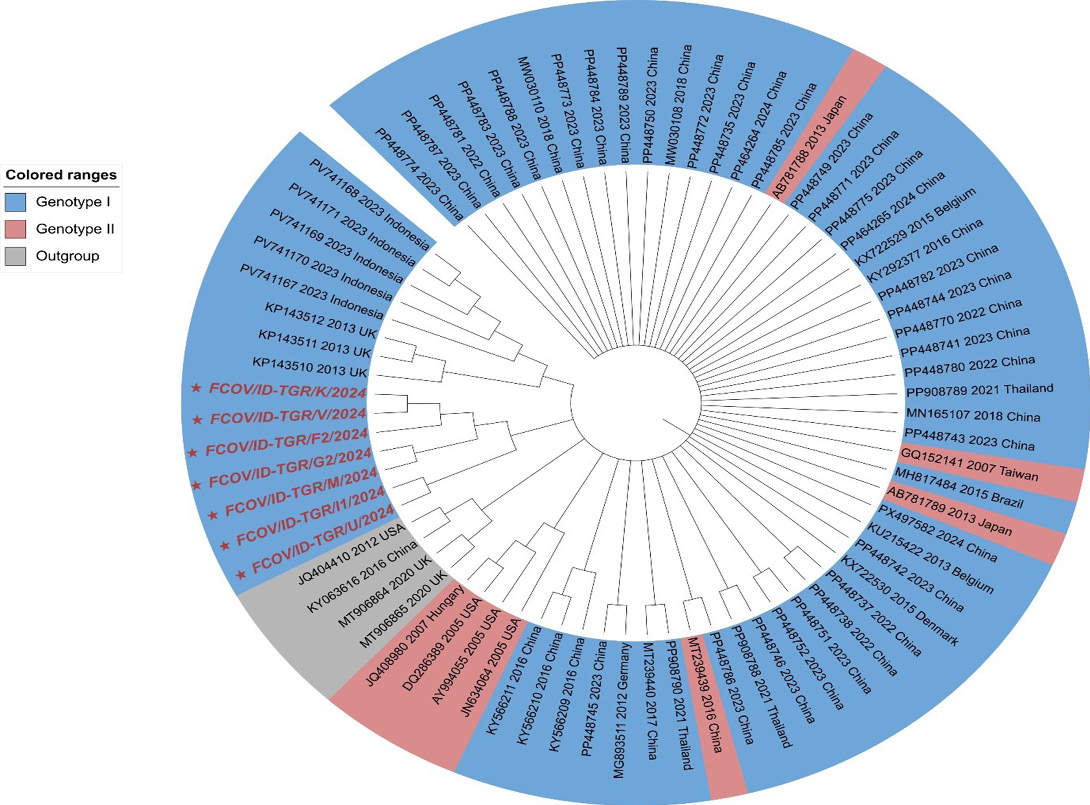
Separate phylogenetic trees were constructed based on the partial N and M gene sequences using the neighbor-joining method with bootstrap analysis. In the partial N
gene phylogenetic tree, the seven FCoV sequences from Tangerang were positioned among reference FCoV sequences from different geographical origins, including the United Kingdom, USA, China, and Indonesia (Figure 3).
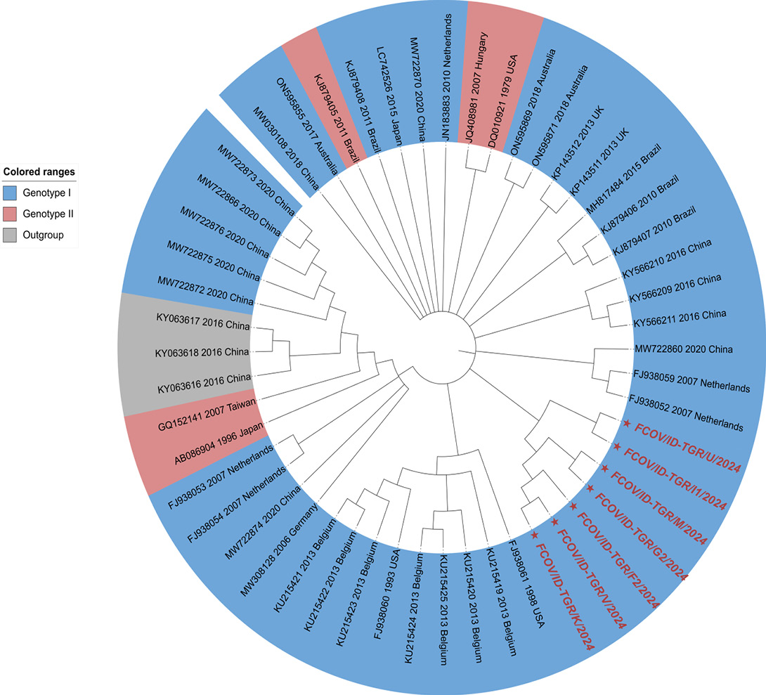
Similarly, phylogenetic analysis based on the complete trimmed M gene sequences (768–771 bp) showed that the seven Tangerang FCoV sequences were distributed among reference sequences from various geographical origins, including China, Belgium, the USA, and the Netherlands (Figure 4). Reconstruction of the M gene phylogenetic tree using the Kimura 2-parameter model was completed with full bootstrap support across all 1,000 replicates. The Tangerang sequences are indicated by star symbols (★) and red font in Figure 4.
DISCUSSION
The molecular detection of Feline coronavirus (FCoV) in this study utilized reverse transcription-polymerase chain reaction (RT-PCR), this approach is highly effective for confirming the presence of viral genetic material, though its sensitivity can be influenced by viral load compared to nested RT-PCR or RT-qPCR .
From 11 archived samples which were previously found to be FCoV-positive based on 3′ UTR and 10 of those samples that were confirmed to be FCoV-positive based on the spike (S) gene , seven samples (63.6%) were successfully amplified based on the nucleocapsid (N) and membrane (M) genes (Table 2). The presence of specific bands at 1202 bp for the N gene and 880 bp for the M gene (Figures 1 and 2), indicating successful amplification of products corresponding to the expected target sizes ; . The identity of the amplified products as FCoV N and M gene sequences was further supported by sequencing and BLASTn analysis, which demonstrated moderate nucleotide identity with FCoV reference sequences (Table 2). While most positive samples showed robust DNA bands, sample K initially exhibited a faint band for the M gene, likely due to low target concentration or minor inhibitors (Figure 2). The electrophoresis was therefore repeated to verify the consistency of the observed amplification result.
The failure to amplify the N and M genes in four samples (D, L, O1, and S), despite their previous positivity for the S gene, may be associated with a low concentration or reduced integrity of viral RNA in the archived samples. Long-term storage, particularly of fecal samples, may reduce viral RNA integrity compared with effusion samples . The double bands observed during M gene amplification most likely indicate the presence of non-specific or multiple amplification products. However, the exact nature of these products could not be determined because they were not separately excised and sequenced. Similarly, overlapping or secondary peaks in some Sanger sequencing chromatograms may have resulted from mixed amplification products or sequencing-related artifacts. These overlapping signals can complicate sequence interpretation and reduce the usable sequence length after quality trimming .
BLASTn analysis showed that the N gene shared 91.22–94.23% nucleotide identity with the Chinese genotype I reference strain PV246293, whereas the M gene exhibited 92.63–94.43% identity with the Chinese genotype I reference strain PP444763. While these values indicate moderate sequence divergence from reference strains, species demarcation within the family Coronaviridae is formally defined by amino acid identity across conserved replicase domains rather than nucleotide identity in individual structural genes (ICTV, 2025).
Within individual strains, the M gene consistently exhibited slightly higher nucleotide conservation compared to the N gene. This difference may reflect distinct gene-specific evolutionary constraints and structural roles. The coronavirus membrane (M) protein is an essential structural component required for virion assembly, envelope formation, and interactions with other viral proteins, such as the spike protein ; . These vital biological functions likely impose functional constraints that limit nucleotide mutation accumulation. In contrast, the nucleocapsid (N) protein primarily functions in binding and packaging viral RNA, virion assembly, and host–virus interactions ; , which may allow for greater sequence variability.
Phylogenetic analysis based on the N gene revealed that all seven Tangerang strains (marked ★ in red font) formed a distinct monophyletic clade, suggesting localized genetic evolution (Figure 3). In contrast, the M gene tree resolved the Tangerang sequences into two closely related lineages within Genotype I: one comprising strains U and I1 clustering near European reference strains, and another containing the remaining five Tangerang isolates (Figure 4). Despite these minor topologies, sequences in both targets exhibited close relationships with reference strains from diverse geographical regions, including China, Europe, and the USA. The genetic relatedness between Indonesian and foreign strains does not establish the route of viral dispersal. While international cat movement has been proposed for other feline pathogens , robust epidemiological and genomic epidemiological data are needed to confirm such routes for FCoV.
The N gene’s conservative nature was evident as genotype II references did not form a single distinct cluster but were scattered (Figure 3), reflecting the gene’s stability against mutation . However, the high conservation of the N gene in both FCoV genotypes makes the N gene a good target for the development of an FCoV vaccine . The M gene phylogeny also showed a scattered distribution of genotype II reference strains (Figure 4), with some positioned near the CCoV outgroups. This pattern is consistent with the previously proposed recombinant origin of genotype II FCoV involving genotype I FCoV and CCoV , although recombination cannot be confirmed from the M gene phylogeny alone.
This study has several limitations that should be noted. First, the sample size was relatively small and restricted to archived clinical specimens from a single geographic region (Tangerang), which may not fully capture the complete genetic diversity of FCoV across Indonesia. Second, quantitative assessment of viral RNA concentration (e.g., via NanoDrop or RT-qPCR) was not conducted, precluding precise correlation between RT-PCR negative results and viral load. Finally, although Sanger sequencing provided accurate target verification, technical constraints such as chromatogram noise, non-specific amplification, and overlapping sequencing signals limited the usable consensus sequence lengths for some isolates.
CONCLUSION
This study characterized partial membrane (M) and nucleocapsid (N) gene sequences of FCoV from Tangerang, Indonesia. Both genes were successfully amplified and sequenced from seven of the 11 archived FCoV-positive samples. BLASTn and phylogenetic analyses showed genetic relatedness between the Tangerang sequences and genotype I reference strains from different geographical regions. The N gene sequences formed a distinct monophyletic clade, whereas the M gene sequences were resolved into two closely related lineages within genotype I. These findings provide baseline molecular epidemiological information on FCoV circulating in Tangerang and may support continued surveillance and future investigations of its genetic diversity and evolution in Indonesia.
ACKNOWLEDGEMENTS
The authors would like to thank the Ministry of Higher Education, Science, and Technology of the Republic of Indonesia for granting a research fund through the Regular Fundamental Research Scheme, under Contract No. 18810/IT3.D10/PT.01.03/P/B/2026 for the 2026 fiscal year.
NOVELTY STATEMENT
This study provides molecular characterization of the partial membrane (M) and nucleocapsid (N) genes of FCoV circulating in Tangerang, Indonesia, addressing the limited molecular information available for these genes in Indonesian FCoV strains. The combined sequence and phylogenetic analyses provide baseline information on the genetic characteristics and relationships of local FCoV strains within genotype I.
AUTHOR’S CONTRIBUTION
MA: contributed to laboratory work, data analysis, original draft writing, and manuscript writing. SAAH: contributed to laboratory work, data analysis, and original draft writing. ZWKA, DAR, and RN: contributed to mentoring and supervised the protocols, laboratory work, and data analysis. KS, MF: contributed to reviewed, suggested, and revised the original draft writing. ONP: contributed to conceptualization, reviewed protocols, supervised the study, data analysis, and revised the original draft and manuscript.
Generative AI and AI assisted technology statement
The authors declare that no generative AI and AI assisted technology was used in the creation of this manuscript.
Conflict of interest
The authors have declared no conflict of interest related to the writing of this article, whether related to funds, data, patents, or any other aspect related to it, with any party.
Figures
Figure 1:.
Electrophoresis results of RT-PCR FCoV nucleocapsid (N) gene.
Figure 2:.
Electrophoresis results of RT-PCR FCoV membrane (M) gene.
Figure 3:.
Phylogenetic tree based on partial nucleotide sequences of the feline coronavirus (FCoV) nucleocapsid (N) gene. The seven FCoV sequences obtained in this study are indicated by red labels and asterisks (★).
Figure 4:.
Phylogenetic tree based on partial nucleotide sequences of the feline coronavirus (FCoV) membrane (M) gene. The seven FCoV sequences obtained in this study are indicated by red labels and asterisks (★)A.
References
Amalia F, Wasissa M, Salasia SIO, Lestari FB (2025). Detection and genetic analysis of Feline coronavirus in clinically suspected cats. World Vet. J., 15(3): 802–810. https://doi.org/10.54203/scil.2025.wvj82[CrossRef]
Attipa C, Warr AS, Epaminondas D, O’Shea M, Hanton AJ, Fletcher S, Malbon A, Lyraki M, Hammond R, Hardas A, Zanti A, Loukaidou S, Gentil M, Gunn-Moore D, Mazeri S, Tait-Burkard C (2023). Emergence and spread of feline infectious peritonitis due to a highly pathogenic canine Feline recombinant coronavirus. bioRxiv., 11.08.566182. https://doi.org/10.1101/2023.11.08.566182[CrossRef]
Bahoussi AN, Wang PH, Shah PT, Bu H, Wu C, Xing L (2022). Evolutionary plasticity of zoonotic Porcine deltacoronavirus (PDCoV): genetic characteristics and geographic distribution. BMC Vet. Res., 18(1): 444. https://doi.org/10.1186/s12917-022-03554-4[CrossRef]
Brostoff T, Savage HP, Jackson KA, Dutra JC, Fontaine JH, Hartigan-O’Connor DJ, Carney RP, Pesavento PA (2024). Feline infectious peritonitis mRNA vaccine elicits both humoral and cellular immune responses in mice. Vaccines, 12(705): 1–15. https://doi.org/10.3390/vaccines12070705[CrossRef]
Crossley BM, Bai J, Glaser A, Maes R, Porter E, Killian ML, Clement T (2020). Guidelines for Sanger sequencing and molecular assay monitoring. J. Vet. Diagn. Invest., 32(6): 767-775. https://doi.org/10.1177/1040638720905833[CrossRef]
de Haan CAM, Smeets M, Vernooij F, Vennema H, Rottier PJM (1999). Mapping of the coronavirus membrane protein domains involved in interaction with the spike protein. J. Virol., 73(9): 7441–7452. https://doi.org/10.1128/JVI.73.9.7441-7452.1999[CrossRef]
de Haan CAM, Vennema H, Rottier PJM (2000). Assembly of the coronavirus envelope: Homotypic interactions between the M proteins. J. Virol., 74(11): 4967–4978. https://doi.org/10.1128/JVI.74.11.4967-4978.2000[CrossRef]
Delaplace M, Huet H, Gambino A, Le Poder S (2021). Feline coronavirus antivirals: a review. Pathogens, 10: 1150. https://doi.org/10.3390/pathogens10091150[CrossRef]
Dong B, Zhang X, Zhong X, Hu W, Lin Z, Zhang S, Deng H, Lin W (2024). Prevalence of natural Feline coronavirus infection in domestic cats in Fujian, China. Virol. J., 21(2): 1–8. https://doi.org/10.1186/s12985-023-02273-y[CrossRef]
Dong HV, Rapichai W, Rattanasrisomporn A, Rattanasrisomporn J (2025). Feline coronavirus in Northern Vietnam: genetic detection and characterization reveal predominance of type I viruses. Viruses, 17: 188. https://doi.org/10.3390/v17020188[CrossRef]
Fitrawati F, Suwarno, Yudaniayanti IS, Rahmahani J, Yunus M, Soeharsono, Karo-karo D (2025). Haeriah. Molecular detection of FIPV among imported felines through Soekarno Hatta Airport, Indonesia. Media Kedokteran Hewan, 36(1): 40–49. https://doi.org/10.20473/mkh.v36i1.2025.40-49[CrossRef]
He WT, Ji X, He W, Dellicour S, Wang S, Li G, Zhang L, Gilbert M, Zhu H, Xing G, Veit M, Huang Z, Han GZ, Huang Y, Suchard MA, Baele G, Lemey P, Su S (2020). Genomic epidemiology, evolution and transmission dynamics of Porcine deltacoronavirus. Mol. Boil. Evol., 37(9): 2641–2654 .https://doi.org/10.1093/molbev/msaa117[CrossRef]
International Committee Taxonomy Viruses (ICTV) (2025). Family: Coronaviridae. ICTV Report. Available at: https://ictv.global/report/chapter/coronaviridae/coronaviridae (Accessed September 4, 2026).
Kopduang C, Rapichai W, Leangcharoenpong C, Khamsingnok P, Puangmalee T, Ratanabunyong S, Rattanasrisomporn A, Khaoiam T, Dong HV, Choowongkomol K, Rattanasrisomporn J (2025). Detection of Feline coronavirus membrane gene based on conventional revere transcription-polymerase chain reaction, nested reverse transcription-polymerase chain reaction, and reverse transcription-quantitative polymerase chain reaction: a comparative study. Int. J. Mol. Sci., 26: 6861. https://doi.org/10.3390/ijms26146861[CrossRef]
Kumar S, Stecher G, Suleski M, Sanderford M, Sharma S, Tamura K (2024). MEGA12: molecular evolutionary genetic analysis version 12 for adaptive and green computing. Mol. Biol. Evol., 41: 1–9. https://doi.org/10.1093/molbev/msae263[CrossRef]
Le Poder S (2011). Feline and canine coronaviruses: Common genetic and pathobiological features. Adv. Virol., 2011: 609465. https://doi.org/10.1155/2011/609465[CrossRef]
Letunic I, Bork P (2024). Interactive tree of life (iTOL) v6: recent updates to the phylogenetic tree display and annotation tool. Nucleic Acids Res., 52: W78–W8. https://doi.org/10.1093/nar/gkae268[CrossRef]
Li J, Jia H, Tian M, Wu N, Yang X, Qi J, Ren W, Li F, Bian H (2022). SARS-CoV-2 and emerging variants: unmasking structure, function, infection, and immune escape mechanisms. Front. Cell. Infect. Microbiol., 12: 869832. https://doi.org/10.3389/fcimb.2022.869832[CrossRef]
Marzok M, Kandeel M, Al-Mubarak A, Farag A, Elgohary A, El-khodery S (2023). Global prevalence of Feline coronavirus infection (FCoV) in domestic cats: systematic meta-analysis. J. Anim. Plants Sci., 33(6): 1269–1285. https://doi.org/10.36899/JAPS.2023.6.0689[CrossRef]
Meli ML, Spiri AM, Zwicklbauer K, Krentz D, Felten S, Bergmann M, Dorsch R, Matiasek K, Alberer M, Kolberg L, Both V, Hartmann K, Hofmann-Lehmann R (2022). Fecal Feline coronavirus RNA shedding and Spike gene mutations in cats with feline infectious peritonitis treated with GS-441524. Viruses, 14: 1069. https://doi.org/10.3390/v14051069[CrossRef]
Mulyani GT, Budhi S, Raharjo S (2024). Corona Virus in Dogs and Cats in Yogyakarta and Its Surroundings. J. Sain Veteriner, 42(2): 282–286. https://doi.org/10.22146/jsv.83658[CrossRef]
National Center for Biotechnology Information (NCBI) (2025). Feline coronavirus isolate FCoV-5-12 Spike protein (S), non-structural protein 3a (ORF3a), non-structural protein 3b (ORF3b), non-structural protein 3c (ORF3c), Envelope protein (E), Membrane protein (M), Nucleocapsid protein (N), non-structural protein 7a (ORF7a), and non-structural protein 7b (ORF7b) genes, complete cds. Available at: https://www.ncbi.nlm.nih.gov/nuccore/PV246293 (accessed 23 Jul 2026).
Poetri ON, Nadila R, Hidayat R, Jesifa LJ, Alfin ZWK, Handharyani E, Mayasari NLPI, Rahmi VA, Rizaldi DA (2026). Molecular characterization and phylogenetic analysis reveal a distinct genotype I FCoV sub-lineage in Tangerang, West Java, Indonesia. Pak. Vet. J., 46(4): 1047–1050.
Salomonsen, BUH, Mortensen, Halkier, BA (2014). DNA cloning and assembly methods: USER-derived cloning methods and their primer design. Humana Press, USA. https://doi.org/10.1007/978-1-62703-764-8_5[CrossRef]
Shi K, He M, Shi Y, Long F, Shi Y, Yin Y, Pan Y, Li Z, Feng S (2024). Genetic and phylogenetic analysis of Feline coronavirus in Guangxi Province of China from 2021 to 2024. Vet. Sci., 11: 455. https://doi.org/10.3390/vetsci11100455[CrossRef]
Tasker S, Addie DD, Egberink H, Hofmann-Lehmann R, Hosie MJ, Truyen U, Belák S, Boucraut-Baralon C, Frymus T, Lloret A, Marsilio F, Pennisi MG, Thiry E, Möstl K, Hartmann K (2023). Feline infectious peritonitis: european advisory board on cat diseases guidelines. Viruses, 15: 1847. https://doi.org/10.3390/v15091847[CrossRef]
Phylogenetic Analysis of Feline Coronavirus (FCoV) Isolated from Tangerang, Indonesia Based on the Nucleocapsid (N) and Membrane (M) Genes · ResearchersLinks