
Toxoplasmosis is one of the most important zoonotic diseases with serious health risks for wild animals. The South China tiger, the most endangered tiger subspecies endemic to China, remain only a little more than 200 today, was susceptibly infected with Toxoplasma gondii. In this study, we used polymerase chain reaction to detect DNA, and enzyme-linked immunosorbent assay to test for T. gondii antibodies. Antibodies (S/P>0.31) to T. gondii were found in 20 (26.32%) of the 76 tigers, while all blood samples tested through nested PCR were negative. This is the first investigation of T. gondii infection in South China tiger.
Short Communication · Volume 55, Issue 4, 22 July 2023 · Open Access
Molecular and Serological Detection of Toxoplasma gondii in South China Tiger
· ·
Affiliations & Notes ⌄
- 1Shanghai Zoo, 2381 Hongqiao Road, Changning District, Shanghai, China, 200335 * Corresponding author: petitella@163.com
Article Info ⌄
- Journal:
- Pakistan Journal of Zoology
- Published:
- 22 July 2023

From the article
- Antibodies (S/P>0.31) to T. gondii were found in 20(26.32%) of the 76 tigers, sampled from all nine sampling regions: Shanghai, Hangzhou, Chengdu, Chongqing, Guangzhou, Meihuashan, Nanchang, Suzhou and Linyi.
- The difference in the frequency of S/P ratio on T. gondii infection in male (X =0.26, SD=0.16) and female (X =0.22, SD=0.13) tigers was not significant.
- The antibody-positive rate of sub-adult tigers (80.0%) was higher compared with young (16.0%) and adult tigers (26.1%).
- Epidemiological survey results for the detection of T. gondii are available in Table I. The prevalence of T. gondii in the Chongqing and Chengdu tigers was noticeably higher compared to tigers in other places in China.
Selected from the article's own text.
Abstract
Materials and methods
Seventy blood samples of captive South China tigers were collected from nine zoos (Shanghai, Hangzhou, Chengdu, Chongqing, Guangzhou, Meihuashan, Linyi, Suzhou, Nanchang) in China (Fig. 1). Sera were separated by centrifugation at 2,000 g for 5 min. The sera were stored at -20°C for toxoplasma antibodies detection, and the blood samples were processed for genomic DNA extraction using a Wizard® Genomic DNA Purification Kit (Promega, Madison, WI, USA), according to the manufacturer’s instructions.
T. gondii was detected by Nested PCR targeting B1 DNA around the XhoI and PmlI restriction sites, according to a previously described method . PCR primers used for amplification are as follows: Pml/S1, 59-TGTTCTGTCCTATCGCAACG (positions 128 to147); Pml/S2, 59-TCTTCCCAGACGTGGATTTC (positions 152 to 171); Pml/AS1, 59-ACGGATGCAGTTCCTTTCTG (positions 688 to 707); and Pml/AS2, 59-CTCGACAATACGCTGCTTGA (positions 663 to 682), yielding a 504bp product. PCR was carried out in the final volume of 20μL containing 10μLPCR mix (2×) (Takara Dalian, China), 0.4 μL of each forward and reverse primer (10μmol/L), 2μL DNA (200 ng/mL) and 7.2 μL DNase/RNase-free water. The amplified PCR products were separated by electrophoresis in 1.5% agarose gels.
Serum antibodies against T. gondii were screened using the EVL Toxo test ELISA kit (European Veterinary Laboratory, Woerden, The Netherlands), according to the manufacturer’s recommendations. The serum samples and controls were diluted to 1:500 and tested in duplicate. The optical density (OD) was measured at 450 nm with an ELISA plate reader (Thermo Fisher, Waltham, MA, USA). The S/P (samples/positive control) ratio for each sample was calculated according to the formula: S/P = (OD450of the sample - OD450 of negative control)/ (OD450ofpositive control - OD450 of negative control). Samples with S/P ratio lower than 0.31 were considered negative for T. gondii antibodies. If the S/P ratios were greater than or equal to 0.31, the samples were considered positive.
The prevalence of T. gondii infection in South China tigers of different sexes, ages, regions, and sampling times was analyzed using the Chi Square Test in SPSS (version 18.0, SPSS Inc. Chicago, IL, USA). A probability (p) value of< 0.05 was considered statistically significant.
Results and discussion
The present study is the first epidemiological investigation of T. gondii infection in South China tiger in China. Antibodies (S/P>0.31) to T. gondii were found in 20(26.32%) of the 76 tigers, sampled from all nine sampling regions: Shanghai, Hangzhou, Chengdu, Chongqing, Guangzhou, Meihuashan, Nanchang, Suzhou and Linyi. The seropositive rates in Chongqing (66.7%) and Chengdu (50.0%) were significantly higher than those in the other sampling regions. The difference in the frequency of S/P ratio on T. gondii infection in male (X =0.26, SD=0.16) and female (X =0.22, SD=0.13) tigers was not significant. The antibody-positive rate of sub-adult tigers (80.0%) was higher compared with young (16.0%) and adult tigers (26.1%). Sampling time had an impact on the results the positive rate of serum samples was much higher in October and December than in other months (January, February, April, July, November). Variance in test results on the basis of region, age, or time of the sample for the detected parasite showed significant differences. Epidemiological survey results for the detection of T. gondii are available in Table I.
The prevalence of T. gondii in the Chongqing and Chengdu tigers was noticeably higher compared to tigers in other places in China. This may be related to sporulation and survival of coccidial oocysts in the environment due to temperature and humidity.
We found that the offspring of antibody-positive female tigers were also antibody-positive. This is consistent with the conclusion of Sharma et al. (2019). T. gondii infection can transmit tachyzoites from mother to fetus through vertical transmission .
Table I. General characteristics and prevalence of Toxoplasma gondiiin South China tigers in Eastern China (n=76).
| Variables | Antibody level | Seropositive | Nested PCR | |||||
| N | Mean±SD | 95% CI | P value | % ELISA | n(%) | |||
| Lower limit | Upper limit | |||||||
| Region | ||||||||
| Shanghai | 60 | 0.29±0.159 | 0.21 | 0.36 | 0.01 | 2(3.33) | 0 | |
| Hangzhou | 13 | 0.27±0.12 | 0.20 | 0.34 | 3(23.08) | 0 | ||
| Chengdu | 4 | 0.29±0.16 | 0.03 | 0.544 | 2(50.00) | 0 | ||
| Chongqing | 6 | 0.359±0.18 | 0.16 | 0.55 | 4(66.67) | 0 | ||
| Guangzhou | 9 | 0.269±0.13 | 0.16 | 0.36 | 4(44.44) | 0 | ||
| Meihuashan | 6 | 0.109±0.16 | -0.07 | 0.27 | 1(16.67) | 0 | ||
| Linyi | 2 | 0.12±0.00 | 0.11 | 0.13 | 0(0) | 0 | ||
| Suzhou | 1 | -0.04±\ | \ | \ | 0(0) | 0 | ||
| Nanchang | 15 | 0.19±0.09 | 0.13 | 0.24 | 1(6.67) | 0 | ||
| Gender | ||||||||
| Male | 40 | 0.26±0.16 | 0.21 | 0.32 | 0.19 | 13(32.50) | 0 | |
| Female | 36 | 0.22±0.13 | 0.18 | 0.26 | 7(19.44) | 0 | ||
| Age | ||||||||
| ≤1 year | 25 | 0.20±0.18 | 0.13 | 0.28 | 0.02 | 4(16.00) | 0 | |
| 1-3 year | 5 | 0.41±0.10 | 0.28 | 0.54 | 4(80.00) | 0 | ||
| >3 year | 46 | 0.24±0.13 | 0.20 | 0.28 | 12(26.09) | 0 | ||
| Time | ||||||||
| January | 6 | 0.10±0.16 | -0.07 | 0.28 | 0.01 | 1(16.67) | 0 | |
| February | 2 | 0.12±0.00 | 0.11 | 0.14 | 0(0) | 0 | ||
| April | 1 | 0.21±\ | \ | \ | 0(0) | 0 | ||
| July | 6 | 0.15±0.12 | 0.02 | 0.28 | 0(0) | 0 | ||
| October | 19 | 0.30±0.13 | 0.24 | 0.37 | 6(31.58) | 0 | ||
| November | 25 | 0.22±0.12 | 0.17 | 0.27 | 4(16.00) | 0 | ||
| December | 17 | 0.31±0.15 | 0.23 | 0.39 | 9(52.94) | 0 | ||
| Total | 76 | 0.24±0.15 | 0.21 | 0.28 | 20(26.32) | 0 | ||
T. gondii can infect a variety of mammals. Though felines are the primary hosts for the parasite, other animals feed on the egg sacs of felines and become infected ; . At present, captive African lions (Panthera leo), Lynxes (Lynx canadensis), and Siberian tigers (Panthera tigris altaica) are susceptible to T. gondii . Evidence of infection mostly discovered through serological detection has been found for 31 of the world’s 39 felid species .
It is necessary to prevent and control the spread of T. gondii during daily feeding. First, we should control the number of stray cats, optimize the layout of zoos, and reduce contact between stray cats and South China tigers. Additional measures will include strengthening disinfection management, interrupting the transmission route of T. gondii, and ensuring the health and safety of infected South China tigers by quarantining them even after treatment, which in turn will guard the health of other wild animals in the zoo. Preventing cross infection and repeat infection in zoos is crucial in managing this dangerous condition.
In our study, all blood samples tested through nested PCR were negative for the presence of T. gondii. Lack of positive results from nested PCR in seropositive tigers was most likely related to T. gondii infection in tigers in the past. Moreover, the difference between the results may derivefrom the fact that T. gondii is temporarily present in blood, and it is therefore possible that it is not readily detected by nested PCR.
Acknowledgements
The authors would like to thank the staff of Shanghai Zoo, Hangzhou Zoo, Chengdu Zoo, Chongqing Zoo, Guangzhou Zoo, Longyan Meihuashan Nature Reserve, Nanchang Zoo, Suzhou Zoo and Linyi Zoo, for assistance in handling the tigers.
Funding
This research was supported by he Special Fund for Scientific Research of Shanghai Landscaping & City Appearance Administrative Bureau (GZ200404); the Opening Foundation of Beijing Key Laboratory of Captive Wildlife Technologies in Beijing Zoo (ZDK202102); and the Science and Technology Project of Shanghai Zoo (SZ170305; SZ190204; SZ160206).
Statement of conflict of interest
The authors have declared no conflicts of interest.
Figures
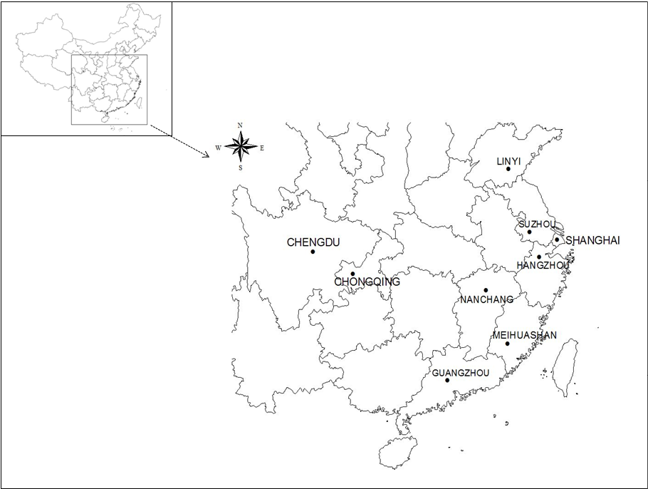
Map of surveyed cities in China, where the blood samples of tigers were collected. The black dots indicate the sample collection areas.
References
- Chen, R.F., Chen, X.L., and Tang, Y., 2014. Anim. Husband. Vet., 46: 25-28.
- Cutler, S.J., Fooks, A.R., and van der Poel, W.H., 2010. Emerg. Infect. Dis., 16: 1-7. https://doi.org/10.3201/eid1601.081467[CrossRef]
- Daszak, P., Cunningham, A.A., and Hyatt, A.D., 2000. Science, 287: 443-449. https://doi.org/10.1126/science.287.5452.443[CrossRef]
- Dubey, J.P., 2008. J. Eukaryot. Microbiol., 55: 467-475. https://doi.org/10.1111/j.1550-7408.2008.00345.x[CrossRef]
- Dubey, J.P., 2016. Toxoplasmosis of animals and humans (Second edition). FL: CRC Press, Inc., Boca Raton.
- Dubey, J.P., and Beattie, C., 1988. Toxoplasmosis of animals and man. CRC Press, Inc., Boca Raton, FL.
- Dubey, J.P., and Su, C., 2009. Mem. Inst. Oswaldo Cruz., 104: 190-195. https://doi.org/10.1590/S0074-02762009000200011[CrossRef]
- Grigg, M.E., and Boothroyd, J.C., 2001. J. clin. Microbiol., 39: 398-400. https://doi.org/10.1128/JCM.39.1.398-400.2001[CrossRef]
- King, A., Varughese, P., De Serres, G., Tipples, G.A., and Waters, J., 2004. J. Infect. Dis., 189 (Suppl 1): S236-242. https://doi.org/10.1086/378499[CrossRef]
- Munhoz, A.D., Hage, S.B., Cruz, R.D.S., Calazans, A.P.F., Silva, F.L., Albuquerque, G.R., and Lacerda, L.C., 2017. Vet. Parasitol. Reg. Stud. Rep., 8: 35-38. https://doi.org/10.1016/j.vprsr.2017.01.007[CrossRef]
- Sharma, R., Parker, S., Al-Adhami, B., Bachand, N., and Jenkins, E., 2019. Fd. Waterb. Parasitol., 15: e00046. https://doi.org/10.1016/j.fawpar.2019.e00046[CrossRef]
- Williams, E.S., Yuill, T., Artois, M., Fischer, J., and Haigh, S.A., 2002. Rev. Sci. Tech., 21: 139-157. https://doi.org/10.20506/rst.21.1.1327[CrossRef]
- Yang, H.Q., 2020. Anim. Poult. Breed. China, 5: 40-41.
Services
Article usage statistics
Loading…