The prevalence rate of T. gondii in pregnant women’s blood samples, detected using the serological rapid test, varied across age groups with significant differences (p ≤ 0.01).
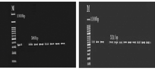
When using polymerase chain reaction (PCR) to amplify the B1 gene regions (580 bp and 531 bp), the infection prevalence in pregnant women was found to be 40%, as shown in (Figure 1).
The PCR results for ruminants revealed an infection prevalence of 10%, with the highest (p ≤ 0.01) prevalence in sheep at 16%, and in goats at 4% (Table 2).
This study concludes that nested PCR is more effective at detecting the B1 gene of T. gondii, facilitating the identification of the agent responsible for abortions in pregnant women.
Selected from the article's own text.
Abstract
Toxoplasma gondii is an essential intracellular parasite responsible for causing toxoplasmosis infections in both humans and animals, which can result in a wide range of clinical signs. In this study, a random blood sample of 165 pregnant women was used for serological testing and molecular analysis. Additionally, 100 serum samples from small ruminants (sheep and goats) were collected from slaughterhouses during the same study period. The prevalence rate in women varied by age categories, with significant differences (p ≤ 0.01): 32.7% in 18–25-year-olds, 21.8% in 25–35-year-olds, and 10.9% in 35–40-year-olds. The B1 gene was detected in both pregnant women and small ruminants using nested PCR. Of the 100 pregnant women, 40 (40%) tested positive using nested-PCR, which amplified 580 bp and 531 bp bands with external and internal sets of primers specific for the B1 gene of T. gondii. In ruminants, PCR revealed a 10% infection rate (10/100), with higher prevalence in sheep (16%, 8/50) compared to goats (4%, 2/50). These findings suggest that the low prevalence of toxoplasmosis infection in ruminants plays a significant role in protecting humans from parasite infections.
Toxoplasma gondii (T. gondii) is one of the most common parasites that infect humans. After the initial infection, immune-compromised patients rely on cell-mediated immunity to help eradicate the infection ; . During pregnancy, a mother may contract toxoplasmosis, which puts the unborn child at risk for serious health issues such as stillbirth, miscarriage, or congenital disabilities. According to Pinto-Ferreira’s research, if the woman has a primary infection, the unborn child is most likely to be infected during the third trimester of pregnancy, with more than 211 million births occurring each year ; . The most common way for toxoplasmosis to spread among the general public is through the ingestion of oocysts, which are shed in the feces of infected cats. Toxoplasmosis can be vertically transmitted from mother to child during pregnancy, with the third trimester being the most common period for transmission. When an acute T. gondii infection occurs, transplacental transmission has been linked to a range of unfavorable outcomes, including stillbirths, congenital abnormalities, and neonatal mortality in mothers with impaired immune systems . Possible outcomes include coma, systemic infection, pneumonia, chorioretinitis, local and systemic brain damage, and even death.
Although humans and cats are the primary reservoirs of the disease, the prevalence of T. gondii in the population of the WHO Eastern Mediterranean Region remains unclear . Immunocompromised patients can eliminate T. gondii infection after the initial infection via cell-mediated immunity. T. gondii can encyst in various organs of a host, including the brain, skeletal muscle, liver, kidney, and heart, with a strong preference for nerve cells, which allows the parasite to form cysts in the brain. Pregnant women are advised to avoid contact with cat litter and soil, as they may contain T. gondii oocysts, which pose a significant risk of infection ; . Congenital toxoplasmosis occurs when the parasite is transmitted from mother to child during pregnancy , leading to serious health concerns for the unborn child. Numerous animal species, including wild, domesticated, and companion animals, can also be infected with T. gondii. Cat feces containing T. gondii oocysts pollute the environment, and the oocysts can sporulate in both terrestrial and aquatic habitats. Toxoplasma infection outbreaks in humans and animals are largely caused by contaminated water reservoirs, as the oocysts are resistant to many chemicals and disinfectants commonly used in the water supply industry .This study aims to determine the susceptibility of pregnant women and small ruminants to T. gondii infection and to detect the parasite using molecular analysis, as opposed to conventional methods.
MATERIALS AND METHODS
Ethical Approval
The current study was approved by the Iraqi Human and Animal Welfare Code at the University of Fallujah, College of Applied Science.
Sample Collection
A total of 165 blood samples from pregnant women were randomly collected between January 2024 and October 2024 in the Al-Anbar province (from Al-Ramadi Gynecology and Pediatrics Educational Hospital and Al-Fallujah Educational Hospital of Gynecology and Pediatrics). These samples were then examined using serological tests and molecular analysis. Additionally, during the same study period, 100 serum samples from small ruminants (goats and sheep) were collected from slaughterhouses.
Serological Diagnosis
Rapid test as directed by the manufacturer (Humatex-Toxo, Human-Diagnostics Worldwide, Germany) was utilized as a screening test for serological confirmation of T. gondii.
DNA Extraction
Following the manufacturer instructions, DNA was extracted from whole blood using a DNA extraction kit (Bioneer, Korea).
Detection of T. Gondii DNA by PCR Assay
Following the manufacturer’s instructions, the genomic DNA was extracted from the blood of the human and small ruminant with T. gondii, the samples of DNA were kept at -20°C. The PCR reaction were conducted by using two specific primers to detect the T. gondii forward primer (5-TGT TCT GTC CTA TCG CAA CG-3), and reverse primer (5-ACG GAT GCA GTT CCT TTC TG-3), forward primer (5-TCT TCC CAG ACG TGG ATT TC-3) and reverse primer (5-CTC GAC AAT ACG CTG CTT GA-3), which amplify 580 bp and 531bp respectively, region of B1 gene . Twenty four microliters was the actual volume used for the PCR reactions, which included 12.5 microliters of green master mix (Promega, USA), 5 microliters of genomic DNA, 1 microliter of each primer, and 5.5 microliters of nucleus-free water. Following a 2-minute initial denaturation stage at 94°C, there were 40 cycles that lasted 1 minute each at 94, 57, and 72 degrees celsius, with a 5-minute final extension stage at 72 degrees Celsius. After the amplification phase, every PCR result was examined. Five liters of the amplified samples were placed right away onto a 1.5% agarose gel electrophoresis and a UV transilluminator.
Statistical Analysis
In order to determine mean differences, the collected data was statistically examined using the Analysis of Variance (ANOVA) and the Chi-square test for categorical variables. The analysis was performed using SPSS software .
Table 1: T. gondii distribution based on rapid test among various age groups of pregnant women.
Age
Positive No.
%
Negative No.
%
Total No.
18-25 years
18
32.7
37
67.2
55
25-35 years
12
21.8
43
78.1
55
35-40 years
6
10.9
49
89
55
Chi-Square (χ2) (P≤0.01) Significant different.
RESULTS AND DISCUSSIONS
The prevalence rate of T. gondii in pregnant women’s blood samples, detected using the serological rapid test, varied across age groups with significant differences (p ≤ 0.01). The rates were 32.7% in those aged 18–25, 21.8% in those aged 25–35, and 10.9% in those aged 35–40 (Table 1). When using polymerase chain reaction (PCR) to amplify the B1 gene regions (580 bp and 531 bp), the infection prevalence in pregnant women was found to be 40%, as shown in (Figure 1). The PCR results for ruminants revealed an infection prevalence of 10%, with the highest (p ≤ 0.01) prevalence in sheep at 16%, and in goats at 4% (Table 2).
Table 2: The prevalence of T. gondii distribution based on the PCR test among goats and sheep.
Host
Total sample No.
Positive No.
%
Sheep
50
8
16
Goat
50
2
4
Total
100
10
10
Chi-Square (χ2) (P≤0.01) Significant different.
The study focused on toxoplasmosis, a foodborne infection that may impact pregnant women. The results of this study disagreement with other study in Bénin, using a rapid test the seroprevalence of toxoplasmosis was 48.9% in older women and in Nigeria, the toxoplasmosis seroprevalence based the result of the rapid test for toxo IgG-IgM was 28.1% . In comparison to study conducted in the Al-Qadisiyah province incompatible with this study, the rate of infection was16% of age groups 20 to 25 and in Kaduna State Northwest Nigeria, ages 16-20 and 26-30 years have the largest seropositive of 0.8%, while ages 21-25 and 31-35 years have a seroprevalence of 0.6%, and all ages 36-40 years are negative ; . Previous study ELISA IgG and IgM tests revealed 34.01% of total positives, which is regarded as a confirmed case of toxoplasmosis. Age group 20–29 years old had the highest rate of IgG 26.66%, whereas age group 30-39 years old had the highest rate of IgM 10.52% . Regarding to PCR results, the result of this study is not consistent with the study in Babylon Province, where it was recorded 19 out of 112(16.9%) was positive to toxoplasmosis by nPCR . In Iran, based on the Toxoplasma SAG1 gene in the blood, 36 (97.3%) cases were positive; in India, for the B1 gene, 13 (8.6%) out of 150 patients were qPCR positive; and in Egypt, the infection rate was 57.3% in aborted women ; ; . Toxoplasmosis has been confirmed by PCR analysis, which detected the B1 gene in the peripheral blood of 9 (90%) out of 10 positive samples and based on the nested-PCR method, T. gondii DNA was detected in 14.18%. It appears that the sample collection type, sampling technique, and other criteria, such as dietary habits, climatic conditions can influence toxoplasmosis prevalence in different places. The previous research on the seroprevalence of toxoplasmosis in pregnant women in Kosovo found that 1.2% of women contracted toxoplasmosis during their pregnancy ; . The results of this study, agreement with another study in Al-Diwaniyah City, Iraq, showed that T. gondii were detected in 40% of women, whereas disagreement in Al-Najaf province revealed that 87.5% were positive for B1 gene detection ; . RT-qPCR was used to identify 14.3% of pregnant women in Maysan Province and PCR was used to identify 32 (51.61%) of pregnant women in Mosul Province who were infected with T. gondii ; . The results of this study disagreement with previous study in India, using a B1 gene based PCR to find T. gondii, sheep tested positive 3.67 while the goat infection rate was 3.50% according to the results . In Iran, the results revealed that T. gondii was present in 26 (5.9%) of the samples . In Baghdad, with regard to the B1 gene the PCR analysis overall positive results indicated that 13.92% in goat and in Al-Qadisiyha, using RT-PCR to identify the particular parasite B1 gene in blood samples revealed 9% sheep with positive infection results and in Duhok, recorded 21.6% in ewe ; . It has been suggested that the low PCR positive rate caused by the parasite not being present in the blood at the time of collection and having already localized as tissue cysts, tachyzoites, or bradyzoites within the host’s body. Additionally, the cause can be that the acute disease goes away or becomes a chronic infection. The variations in positive infections can be attributed to variations in the amount of samples analyzed in the study, as well as uncontrolled laboratory circumstances that can lead to errors in PCR results ; .
CONCLUSIONS AND RECOMMENDATIONS
This study concludes that nested PCR is more effective at detecting the B1 gene of T. gondii, facilitating the identification of the agent responsible for abortions in pregnant women. Additionally, our findings suggest a higher infection rate among young women, likely influenced by various factors, including residency and seasonal variations that impact parasite distribution. Finally, negative PCR results may be due to inhibitors interfering with the DNA extraction process. Comparatively, research on ruminants shows a low infection rate of toxoplasmosis, which plays a significant role in protecting humans from parasite infection.
ACKNOWLEDGEMENTS
The authors wish to acknowledge the field technicians who have assisted in research activity, and the authors would like to thank the Al-Turath University, College of Pharmacy.
NOVELTY STATEMENTS
Our study was carried out honorably, in compliance with the strictest ethical guidelines and relevant laws.
AUTHOR’S CONTRIBUTIONS
Z.K. Al-Ani and S.M. Farhan used statistical analysis to construct the idea, and S.A. Shwan and A.K. Kadhim conducted the laboratory analysis. The final data was written and approved by both authors.
Conflict of Interest
The author should declare any conflict of interest.
Figures
Figure 1:.
A: The PCR results showed amplification of a 580 bp fragment on a 1.5% agarose gel electrophoresis, with a 1000 bp DNA ladder. B: The PCR product revealed amplification of a 531 bp fragment, with a 1000 bp DNA ladder, on a 1.5% agarose gel electrophoresis.
References
Ali S, Amjad Z, Khan TM, Maalik A, Iftikhar A, Khan I and Ahmed H (2020). Occurrence of Toxoplasma gondii antibodies and associated risk factors in women in selected districts of Punjab province, Pakistan. Parasitology, 147(10): 1133-1139. https://doi.org/10.1017/S0031182020000967[CrossRef]
Al-Masoudi HK (2015). Utilization of molecular and serological methods to investigation Toxoplasma gondii in healthy apparently students in Babylon Province. Med. J. Babylon, 12(4):934-42.
Allamy SS, Alsaady HA (2023). Using Antibody rapid test, RT-qPCR, and RFLP/PCR for detecting Toxoplasma gondii in the placenta of abortion women and determining the genotype in Maysan province, Iraq. J. Wasit Sci. Med., 16(3):28-43. https://doi.org/10.31185/jwsm.475[CrossRef]
Al-Omer NS, Al-Marsoomy AH (2021). Molecular and Serological Detection of Toxoplasma gondii in Random Sample of pregnant and aborted women of Nineveh. Coll. Basic Educ. Res. J., 17(2):1848-64. https://doi.org/10.33899/berj.2021.168580[CrossRef]
Amroabadi MA, Rahimi E, Shakerian A (2021). Study of the seasonal and geographical prevalence of Toxoplasma gondii in milk of ruminants by nested-PCR. Food Hygiene. 11:41.
Al-abodi HR (2021). Diagnostic study to detect toxoplasmosis in some Iraqi sheep. J.Phys. Conf. Ser., 1879:2, 022050. https://doi.org/10.1088/1742-6596/1879/2/022050[CrossRef]
Al-Abodi HRJ (2018). Suspicion in the form of infection is the basis for selecting the appropriate method for examining the toxoplasmosis disease of bends that have no symptoms from patients. Int. J. Adv. Res. 6(9):655-662. https://doi.org/10.21474/IJAR01/7721[CrossRef]
Al-Gharibawi YK, Alwaaly AB (2021). Pregnant Women with Toxoplasmosis In Al-Hai City, Wasit Governorate, Iraq. Nveo-Natural Volatiles and Essential Oils Journal NVEO; 5431-40.
Aly NS, Kim HS, Marei YM, Elhamshary AS, Bayoumi IR, Omar RE, Mohammed DA, Miyoshi SI, Rashed GA (2023). Diagnosis of Toxoplasmosis using Surface Antigen Grade 1 detection by ELISA, Nano-Gold ELISA, and PCR in pregnant women. Int. J. Nanomed., 31:1335-45. https://doi.org/10.2147/IJN.S401876[CrossRef]
Alkanaq MN, Shallal ZS, Alrammah HS, Alhadi H (2020). Detection of Toxoplasma gondii in blood and milk of infected goats and pregnant women by rapid test cassette and conventional-PCR methods in AL-Qadisiyah province, Iraq. Int. J. Pharm. Res., 12(2). https://doi.org/10.31838/ijpr/2020.12.02.292[CrossRef]
Boothroyd JC (2009). Toxoplasma gondii: 25 years and 25 major advances for the field. Int. J. Parasitol., 39(8), pp.935-946. https://doi.org/10.1016/j.ijpara.2009.02.003[CrossRef]
Cerutti A, Blanchard N, Besteiro S (2020). The bradyzoite: a key developmental stage for the persistence and pathogenesis of toxoplasmosis. Pathogens, 9(3):234. https://doi.org/10.3390/pathogens9030234[CrossRef]
Dasa TT, Geta TG, Yalew AZ, Abebe RM, Kele HU (2021). Toxoplasmosis infection among pregnant women in Africa: A systematic review and meta-analysis. PloS one, 16(7): e0254209. https://doi.org/10.1371/journal.pone.0254209[CrossRef]
Dubey JP (2009). History of the discovery of the life cycle of Toxoplasma gondii. Int. J. Parasitol., 39(8):877-82. https://doi.org/10.1016/j.ijpara.2009.01.005[CrossRef]
Dani A, Al Quraan E (2023). Investigating research students’ perceptions about statistics and its impact on their choice of research approach. Heliyon, 9(10). https://doi.org/10.1016/j.heliyon.2023.e20423[CrossRef]
Datta P, Garg P, Rattan D, Bagga R, Rohilla M, Khurana S, Sehgal R (2024). Comparison of B1 and RE 529 gene targets by real time PCR and LAMP assay for diagnosis of toxoplasmosis in pregnant females. Indian J. Med. Microbiol., 47:100481. https://doi.org/10.1016/j.ijmmb.2023.100481[CrossRef]
Dentico P, Volpe A, Putoto G, Ramadani N, Bertinato L, Berisha M, Schinaia N, Quaglio G, Maggi P (2011). Toxoplasmosis in Kosovo pregnant women. Microbiol. Q. J. Microbiol. Sci., 34(2):203.
Edward IU, Ibrahim G, Kabiru M, Ikeh EI, Shallangwa IB, Edward DS (2024). Detection of B1 Gene in Toxoplasmosis using PCR among Pregnant Women Attending Antenatal Clinic in Kaduna State Northwest Nigeria. S. Asian J. Parasitol., 7(1): 1-11.
Elaadli H, Abo El-Makarem H, Abd Elrahma AH, Shaapan RM, Bessat M (2023). Prevalence and associated risk factors of Toxoplasma gondii infection in sheep and aborted women in Egypt. Iraqi J. Vet. Sci., 37(2): 437-45. https://doi.org/10.33899/ijvs.2022.135777.2518[CrossRef]
Elamin, MH, Al-Olayan EM, Omer SA, Alagaili AN, Mohammed OB (2012). Molecular detection and prevalence of Toxoplasma gondii in pregnant women in Sudan. Afr. J. Microbiol. Res., 6(2): 308-311. https://doi.org/10.5897/AJMR11.1314[CrossRef]
Ghazzay MH, Al-Kelaby KK, Al-Hilli ES, Hadi NR (2020). Diagnosis of toxoplasmosis in pregnant women using enzyme immunoassay and nested real time PCR in Al-Najaf city/Iraq. Int. J. Pharm. Res., 12(2):140-7. https://doi.org/10.31838/ijpr/2020.12.02.0022[CrossRef]
Khan AH, Noordin R (2020). Serological and molecular rapid diagnostic tests for Toxoplasma infection in humans and animals. Eur. J. Clin. Microbiol. Infect. Dis., 39(1):19-30. https://doi.org/10.1007/s10096-019-03680-2[CrossRef]
Kareem HI (2023). Molecular and Serological Detection of The Presence of Toxoplasma gondii in Intermediate Hosts (Women and Sheep) in Al-Diwaniyah City, Iraq. Al-Qadisiyah J. Vet. Med. Sci., 22(1. SupplementI).
Mikaeel FB, Al-Saeed ATM (2020). Serological and molecular diagnosis of Toxoplasma gondii among ewes and horses in duhok province-iraq. Iraqi J. Agric. Sci., 51(4):1212-1219. https://doi.org/10.36103/ijas.v51i4.1100[CrossRef]
Oyeyemi OT, Oyeyemi IT, Adesina IA, Alagaili, AN, Mohammed OB (2020). Toxoplasmosis in pregnancy: a neglected bane but a serious threat in Nigeria. Parasitology, 147(2):127-34. https://doi.org/10.1017/S0031182019001525[CrossRef]
Ogouyèmi-Hounto A, Agbayahoun-Chokki F, Sissinto Savi de Tove Y, Biokou Bankole B, Adinsi de Souza V, Assogba M, Assougbodji A (2014). Evaluation of a rapid diagnostic test in the diagnosis of toxoplasmosis in pregnant women in Cotonou (Bénin). Bull. Soc. Pathol. exotique, 107: 85-89. https://doi.org/10.1007/s13149-014-0355-8[CrossRef]
Raissi V, Taghipour A, Navi Z, Etemadi S, Sohrabi Z, Sohrabi N, Getso M, Shamsaei S, Fasihi Karami M, Raiesi O (2020). Seroprevalence of Toxoplasma gondii and Toxocara spp. infections among pregnant women with and without previous abortions in the west of Iran. J. Obstet. Gynaecol. Res., 46(3):382-8. https://doi.org/10.1111/jog.14184[CrossRef]
Rostami A, Riahi SM, Gamble HR, Fakhri Y, Shiadeh MN, Danesh M, Gasser, RB (2020). Global prevalence of latent toxoplasmosis in pregnant women: a systematic review and meta-analysis. Clin. Microbiol. Infect., 26(6): 673-683. https://doi.org/10.1016/j.cmi.2020.01.008[CrossRef]
Satbige AS, Vijaya Bharathi M, Ganesan PI, Sreekumar C, Rajendran C (2016). Detection of Toxoplasma gondii in small ruminants in Chennai using PCR and modified direct agglutination test. J. Parasitic Dis., 40:1466-9. https://doi.org/10.1007/s12639-015-0713-x[CrossRef]
Saki J, Zamanpour M, Najafian M, Mohammadpour N, Foroutan M (2021). Detection of acute and chronic Toxoplasma gondii infection among women with history of abortion in the Southwest of Iran. J. Parasitol. Res., (1):6693070. https://doi.org/10.1155/2021/6693070[CrossRef]
Tenter AM (2009). Toxoplasma gondii in animals used for human consumption. Mem. Inst. Oswaldo Cruz, 104:364-9. https://doi.org/10.1590/S0074-02762009000200033[CrossRef]