Advances in Animal and Veterinary Sciences
Case Report
Pregnancy Toxemia in a Golden Retriever Bitch, a Case Report
Deniz Nak1, Yavuz Nak1, Abid Hussain Shahzad2*
1Uludag University, Veterinary Faculty, Bursa, Turkey; 2Theriogenology Section, CVAS, Jhang-35200 Pakistan.
Abstract | Pregnancy toxemia is associated with a relative lack or alteration in the carbohydrate metabolism. Ketosis usually occurs during the late gestation in bitches due to inadequate nutrition or inability to eat enough carbohydrates to meet energy demand with increased litter size. A four-year-old, 55 day pregnant Golden Retriever bitch was presented for evaluation of anorexia, weakness, staggering gait, restlessness, tremble, contraction and subsequent coma. Diagnosis of pregnancy toxemia was based on the presence hypoglycemia in blood and ketone bodies in urine. Fetal’s viability was assessed by heart beat using Ultrasonography. The bitch completely recovered after treatment. On 63rd of gestation, whelping of eight healthy pups was observed. Mostly this condition is overlooked by breeders and misdiagnosed by veterinarians, because it is more common in other species of animals, such as small ruminants and Guinea Pig Sows than canine pregnancy. It is potentially life threatening in severe cases and bitches with large litter size are predisposed to pregnancy toxemia particularly Yorkshire terriers and Labradors. Therefore, case is aimed to present clinical signs, diagnosis and treatment of pregnancy toxemia in Golden Retriever Bitch.
Keywords | Pregnancy toxemia, Golden retriever, Hypoglycemia, Hyperketonemia,Whelping
Received | June 15, 2020; Accepted | August 08, 2020; Published | September 01, 2020
*Correspondence | Abid Hussain Shahzad, Theriogenology Section, CVAS, Jhang-35200 Pakistan; Email: ahshahzad@uvas.edu.pk
Citation | Nak D, Nak Y, Shahzad AH (2020). Pregnancy toxemia in a golden retriever bitch, a case report. Adv. Anim. Vet. Sci. 8(11): 1184-1187.
DOI | http://dx.doi.org/10.17582/journal.aavs/2020/8.11.1184.1187
ISSN (Online) | 2307-8316; ISSN (Print) | 2309-3331
Copyright © 2020 Nak et al. This is an open access article distributed under the Creative Commons Attribution License, which permits unrestricted use, distribution, and reproduction in any medium, provided the original work is properly cited.
Introduction
Pregnancy Toxemia (PT) is a common condition of small ruminants at terminal stages of pregnancy carrying multiple fetuses. In large ruminants this condition is more pronounced in beef cattle during the second half of third trimester particularly in cows with twins. Condition is anticipated with reduced caloric intake, low quality fodder at advanced stage of pregnancy coupled with stress. In does with increased fecundity more chances of PT have been found. Over conditioning is also a predisposing cause of this ailment in domesticated animals. Same condition has also been reported in Guinea pig with identical signs and symptoms of small ruminants. There is profound ketonemia and acidosis leading to emaciated, depressed and comatose condition. It is an unusual but documented clinical ailment in canine (Jackson et al., 1980). Bitches carrying a large sized litter in later stages of gestation may find it challenging to eat since compression of the gravid uterus on stomach. As a sequel, bitch is unable to take sufficient caloric intake and to keep pace with increased nutritional demand, body reserves are utilized. This disturbance leads to altered insulin to glucagon proportion. To overcome this deficient energy, fatty acids and glycerol are mobilized under the effect of lipases. These fatty acids and glycerol are used by liver for fetal demand. Under certain instances liver cannot meet the energy demands. Due to break down of fats, excessive ketone bodies are also produced which are toxic and deteriorate the stressed situation during the terminal stages of pregnancy.
These free fatty acids and ketone bodies cannot cross placental barrier in considerable quantity and maternal glucose source remains as sole energy source for developing fetus. Ultimate outcome of this condition has resulted in off-feeding and depressed state. Diagnosis is based on pregnancy status with the number of pups, hypoglycemia and ketonuria. Hypoglycemia is critical and warrants emergency i.v. glucose therapy. If the bitch is on a nutritional plan having carbohydrate contents, relapse is unilaterally otherwise termination of pregnancy is warranted to save the life of the dam. Cesarean section is advised, even pups are not fully matured, if there is life-threat to bitch on emergency basis as a treatment of choice. In early diagnosed cases, forced feeding and glucose inclusion are the best strategies to cope with the situation. In canine diabetic ketosis development is also reported which may be corrected after whelping. These cases respond well after fluid therapy and insulin treatment (Norman et al., 2006). Besides life-threatening condition, frequency of stillbirths is augmented in dogs with PT. Forced-feeding or intravenous feeding may be attempted. Objective of present case is to report the unique and unusual case in small animal practice with unrelated clinical findings in canine as compared to other species.
Case details
A four-year old, Golden Retriever, 55 day pregnant bitch was presented at the Obstetrics clinic of Animal Hospital (Uludag University) for evaluation of anorexia, weakness, staggering gait, restlessness, tremble, contraction and subsequent coma advancing gradually during the last week. Acoording to the history, bitch was being fed with home-made diet and its feeding behaviour was supressed from the past week. This was the second pregnancy. During the preceding pregnancy it whelped three healthy pups. Thorough examination also revealed visual impairment. The bitch was responsive to some extent on forced stimuli.
The TPR was 36.8°C (normal range 37.5-39.2°C), 132 (normal range 200 beats/min during late pregnancy stagges) and 76 (normal range 15-30/min) respectively. Hematologic examination revealed elevated WBC (24.8x10³/μl; range 4-17.6x10³/μl) and decreased RBC (4.25x10⁶/μl; range 4.48-8.53 x10⁹/l), Hb (9.4 g/dl; range 10.5-20.1 g/dl), and hematocrit (27.64%; range 33.6-58.7%). The differential cell count indicated an increased neutrophil (20.7 x 10³/μL; range 2.5–14.3 x 10³/μL). Other hematological findings were within normal range (Plumb, 2015). Serum biochemistry analyses were done by VETSCAN®, Analyzer (Abaxis Inc, USA). This analyzer uses a photometric system for analysis. On the analysis day, samples were removed from storage, thawed under refrigerated conditions and processed. The reagent disk was loaded with 0.1 ml of plasma by using the manufacturer supplied loading pipette and placed into the analyzer according to the manufacturer’s instructions. Biochemical parameters are given in Table 1. The presence of urine ketone (+++/+++) and glucose (-ve) was evaluated using a commercially available urine strip test (Combur 10 test®, Roche Diagnostics) before treatment. Ketone and glucose were negative in urine second day after treatment.
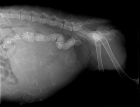
Ultrasonography was done (Siemens Sonoline Prima, Osaka, Japan) with a 5-7.5 MHz linear array transducer to identify the fetal viability or distress. A decreased fetal activity characterized by dorsiflexion of head and limb extension activity was observed. Heart rate of puppies was oberved to be 150-162 (normal 180-240) beats/minute under ultrasound examination done. Lateral and ventrodorsal roentgenograph were obtained to determine the number of puppies. Eight pups were monitored in roentgenography (Figure 1).
Table 1: Comparison of pre and post-treatment biochemical parameters in golden retriever bitch with PT with refrerence values.
| Parameters | Pre treatment | Post treatment | Reference ranges | |
| ALB (g/dL) | 2.90 | 2.90 | 2.60 – 4.0 | |
| ALP(U/L) | 36.0 | 27.0 | 13.0 – 289.0 | |
| ALT(U/L) | 47.0 | 36.0 | 14.0 – 151.0 | |
| AMY(U/L) | 478.0 | 186.0 | 268 – 1653 | |
| TBIL(mg/dL) | 0.30 | 0.30 | 0.10 – 0.50 | |
| BUN(mg/dL) | 5.0 | 11.0 | 8.0 – 30.0 | |
|
CA++(mg/dL) |
8.70 | 7.90 | 8.70 – 12.0 | |
| PHOS(mg/dL) | 2.50 | 2.0 | 2.50 – 7.90 | |
| CREA(mg/dL) | 0.20 | 0.50 | 0.50 – 2.0 | |
| GLU(mg/dL) | 16.0 | 159.0 | 75.0 – 145.0 | |
|
NA+(mmol/L) |
141.0 | 132.0 | 141.0 – 159.0 | |
|
K+(mmol/L) |
5.70 | 4.40 | 3.40 – 5.60 | |
| TP(g/dL) | 6.80 | 7.10 | 5.0 – 8.30 | |
| GLOB(g/dL) | 4.0 | 4.10 | 2.30-5.20 | |
Pregnancy toxemia was diagnosed based on clinical and laboratory findings. First day, 5.0% Dextrose was given 3 times @ 10 ml/kg body weight at 8 hours apart. B-complex @1 ml i.m. Second day, 5.0% Dextrose was given 10 ml/kg SID. There was a quick recovery response after the treatment. Bitch started eating normally and water on second day. After two days observation, the owner was advised to feed with commercially available balanced feed (Royal Canin - Mother and Puppies Starter Medium Dry Food) instead of home-made feed @ free choice to ensure adequate food intake. After initial treatment bitch was evaluated continously and glycaemic codition was noticed till whelping. Following 8th day of treatment the bitch whelped eight haelthy pups without any difficulty.
Discussion
In small ruminants, about 80% fetal development does takes place during the last 45 days of pregnancy and increasing fecundity makes the dam more vulnerable for PT if nutritional deficiency (Bergman, 2015). It has considerable financial burden on small ruminant farming in terms of fetal loss, treatment expenses, and dam mortality. In acute cases, morbidity and mortality rates can reach upto 20% and 80%, respectively (Rook, 2001). Similarly, other small animals like canines there is lack of exact information about this parameter. Fetal heart rate, during canine pregnancy, is a good indicator about the developing fetus well-being. Normal fetal heart rate is 200/min while 180 beats/min indicates stress and <150/minute is an indication of distress necessitating expert intervention (Zone and Wanke, 2001). Although, animals suffering from PT show common disease signs apparently like inapptence, depression, slugishness and marked acetone odor. Coupled with pregnant history these signs are considered as cordinal signs in small ruminats.
The Association of American Feed Control Officials has chalked out minimal feeding requirements for reproduction. This insures sufficient, but not necessarily optimal diet, feed containing 29–32% animal origin protein and 18% fat with balanced omega-6 and omega-3 fatty acids are suggested for pregnant and nursing bitches. In canine pregnancy last two weeks are critical as appetite decreases despite the dams’ body weight gain. In our case there were confused signs of chorea to myoclonus seizures alongwith poor visual perception. Righting reflex was present but jaw champing, head and neck deviation were absent. Reversible nervous signs may be a reason of hypoglycemia which was corrected after proper and prompt treatment. Heart and respiration rates are elevated by 60% and 80% in early and late stages of PT in goats. In canines there is no referral data for this differentiation but history, clinical findings and laboratory aid concludes that present case was at later stages.
Hypoglycaemia and hyperketonaemia both are considered as primary markers for PT (Constable et al., 2017). In present case hypoglycemia was also indicative of alive pups likewise small ruminants (Lima et al., 2012). Reduced serum potassium level in ewes with PT has been described (Halford and Sanson, 1983) on contrary to present case in bitch as shown in table where hayperkalemia was recorded and after treatment its concentration was observed within normal limits. Sodium concentration was found to be at lowest acceptable value and reduced in negative shift post-treatment. From these findings it can be concluded that these levels may be good indicators in small ruminants but not in canines. Plasma calcium and inorganic phosphate concentrations are affected by nutrition. A pronounced decrease in calcium concentration was observed in hyperketonemic ewes in various studies. During the last trimester of pregnancy, the growing fetus also retains an increasing amount of calcium for the circulation, which is required for skeletal development and ewes carrying twins are in even greater need of calcium and are at the same time at a higher risk of developing PT than ewes with single offspring (Hefnawy et al., 2011). In present case both calcium and phosphate concentrations were towards lower limit of normal range.
The bioactivity of liver enzymes increases in response to damage of liver parenchyma and release of these enzymes.In the study carried out by Hefnawy et al. (2011), there was a marked drop in serum total protein, globulin, albumin, cholesterol and total lipids with significant increase in AST and ALT in goats affected by clinical PT which could throw some light on the hepatic origin of PT that may be attributed to fat mobilization associated with inadequate dietary intake or due to hepatic damage or hepatic lipidosis. Range of serum AST and ALT activities in bitch is reported as 14–151U/L and 13–289U/L, respectively.In present case of PT conc. of Albumin, globulin, alkaline phosphates and total proteins were independent of AST. Total bilirubin was also in normal range.
A key feature of pregnancy evaluation status is expectation of prospective troubles. The major mamagemental aim during pregnancy is fetal and maternal well-being. For this monitoring of maternal health status and body condition is of prime importance. In conclusion PT should be always anticipated with large litter size. Owners should be communicated the risk of possible PT and guidlines about feeding managemant must be made available at the time of pregnancy diagnosis.Modern pregnancydiagnosis methods like ultrasonography and roentogram evaluation should be incorporated in routine clinical approaches at beforehand the condition of PT is encountered.Furthermore it is anticipated that unlike various PT markers in small ruminants, in canines hypoglycemia and hyperketonemia are the best indicators for the diagnosis of PT. For fetal well-being assesment, heart rate is another indicator of choice. Present case might be useful for practitioners in their understanding of pathophysiological changes that occur in pregnant bitches during the course of PT.
Authors’ Contributions
DN and AHS designed and wrote the manuscript. Case was handled by DN and AHS. DN and YN performed the ultrasound and hematological evaluation. DN and YN monitored the follow-up.
Conflicts of interest
The authors have declared no conflict of interest.
References


