

Sero-prevalence and Pathological Examination of Lymphoid Leukosis Virus Subgroup A in Chickens in Anhui Province, China


Sero-prevalence and Pathological Examination of Lymphoid Leukosis Virus Subgroup A in Chickens in Anhui Province, China
Hui Zhang2, Yajing Wang2, Kun Li2, Mujeeb Ur Rehman2, Fazul Nabi2, Rui Gui2, Yanfang Lan2 and Houqiang Luo1,2*
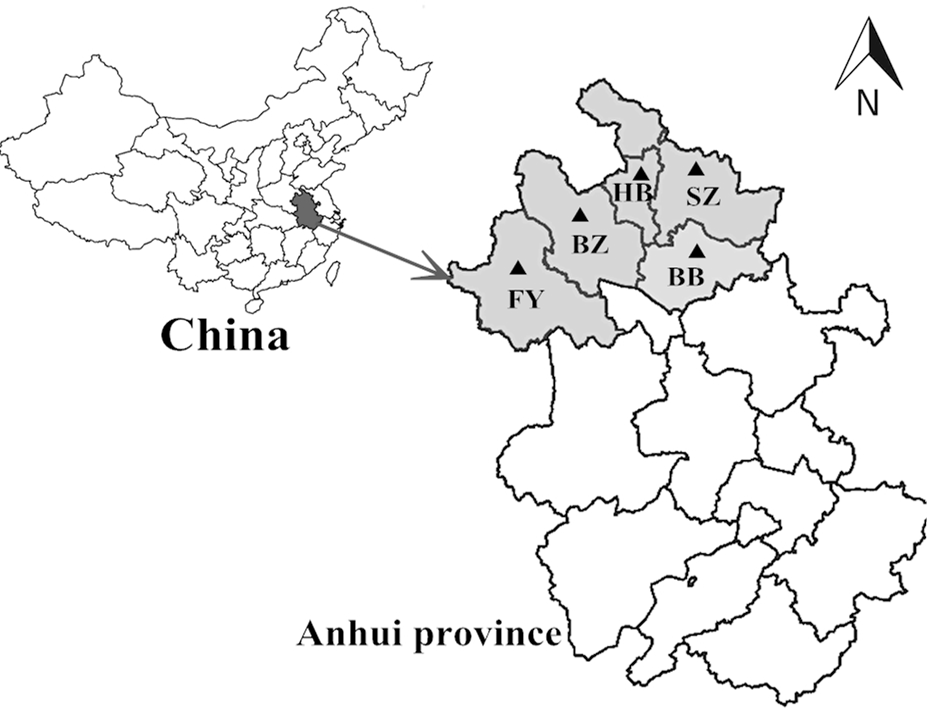
Collection sites of blood samples in Anhui province, China. (HB, Huaibei; SZ, Suzhou; BZ, Bozhou; BB, Bengbu; FY, Fuyang).
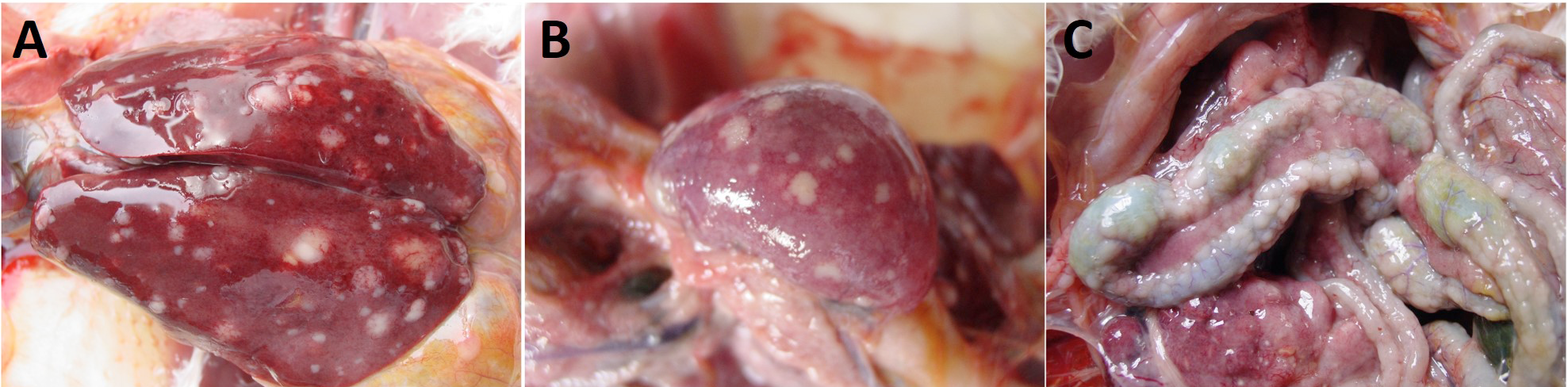
Morphological examination the enlarged liver and diffused nodular tumor lesions with perihepatitis (A), the nodular lesions in the spleen (B) and intestine (C).
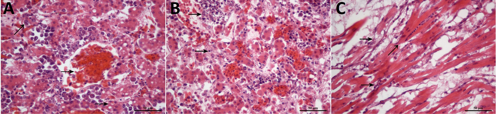
The pathological observation of the congestion in the portal vein with focal lymphoid cells in the liver (A), the degenerative necrosis, hypertrophy with edema on the walls of follicular blood vessels and cells infiltration in the spleen (B) and mild myocardial necrosis, lymphocytic myocarditis, hyperplasia of myocyte nuclei, myocardial infiltration with rupture and myocarditis in the heart (C). Stain, H&E; Magnification, 400x.

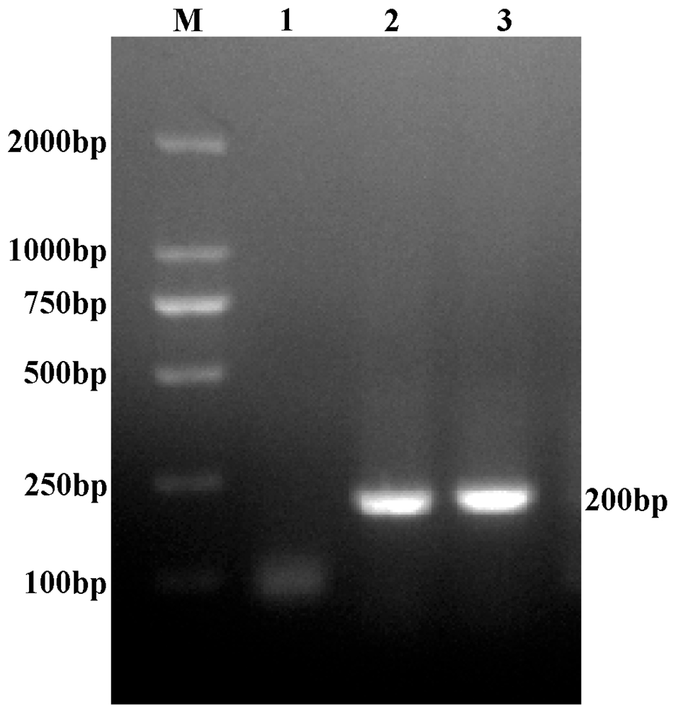
Specific PCR amplification of the ALV-A gene (200bp) on 1.5% agarose gel. M, bp DNA ladder.


