Advances in Animal and Veterinary Sciences
Research Article
Epidemiology of Ticks Fauna of Camels in Samawah Desert
Al Salihi A. Karima1*, Abdulkarim J Karim2, Hussein Jabar Jasim1, Fatima Atiya Kareem1
1College of Veterinary medicine/ Al Muthanna University/ Iraq; 2Faculty of veterinary Medicine/ University of Baghdad, Iraq.
Abstract | Ticks are between the crucial significant vectors of pathogens distressing animals, and also cause health problems like tick paralysis, dermatitis, anemia and secondary infections. This study designed to investigate tick species that infest camels in Al Muthanna province/ Iraq. This study was conducted as a survey of the hard ticks (Acari, Ixodidae) during February 2017-February 2018 on four camels populations (Camelus dromedarius) in different areas in Al Muthanna province. On each occasion, all the visible ticks were collected from the body of each animal. Later on, ticks were transferred to the laboratory, processed and identified microscopically. A total, 1455 ticks were collected during the study period. The overall incidence of the tick’s infestation was 97.1 %. The highest infestation percentage was 98.48 % in female, while it was 75% in male. Ticks were found on different site on the body (legs, chest, axillary, udder, testes, anus, inguinal, ear and face) with more severe lesions on the udder. Hylomma spp. and Boophilus spp. were the most abundant species of ticks found on the camels. In conclusion, this study approved heavy ticks infestation between the camels population in Al Muthanna province accompanied with the variations in the locations and severity of the clinical signs. The authors recommend a future study that contribute to the understanding the species and distribution of ticks that infest camelids in Iraq in order to prevent the implications of ticks infestation and possible prevention measures for diseases transmitted by ticks.
Keywords | Boophilus spp, Camel, Fauna, Hylomma spp. ticks
Editor | Kuldeep Dhama, Indian Veterinary Research Institute, Uttar Pradesh, India.
Received | May 12, 2018; Accepted | June 27, 2018; Published | August 06, 2018
*Correspondence | Al Salihi A. Karima, College of Veterinary medicine/ Al Muthanna University/ Iraq; Email: kama-akool18@mu.edu.iq
Citation | Al-Salihi KA, Karim AJ, Jasim HJ, Kareem FA (2018). Epidemiology of ticks fauna of camels in samawah desert. Adv. Anim. Vet. Sci. 6(8): 311-316.
DOI | http://dx.doi.org/10.17582/journal.aavs/2018/6.8.311.316
ISSN (Online) | 2307-8316; ISSN (Print) | 2309-3331
Copyright © 2018 Al-Salihi et al. This is an open access article distributed under the Creative Commons Attribution License, which permits unrestricted use, distribution, and reproduction in any medium, provided the original work is properly cited.
Introduction
Al Muthanna Province is protected with desert plants and sporadic pastures of varied concentrations (Al Salihi et al., 2017). This severe state is the proper to camels and for this cause they are one of the most important animal resource in Al Muthanna governorate. Merely, the Arabian camels (Camelus dromedarius) are reared in Al Muthanna and they show a very important role in the life of people (as Arabian), they are used as meat, dairy and transportation animals ,beside using as deposited wealth for the forthcoming severe times. Indeed, camels similar other animals are affected with a number of diseases and parasites (Al- Zubaidy, 1995).The external parasites of camels include ticks, mites, and different diseases including parasitic arthropods e.g. myiasis flies (Al- Zubaidy, 1995; Soulsby, 1986). Ticks are a main limit on the world’s livestock production (Zeleke and Bekele, 2004). It employs a major interference to enhance animal production in the tropical and subtropical regions of the world by spreading overwhelming and often deadly livestock diseases, causing blood loss, damage to hides and udder, and paralysis (Dalgliesh et al., 1990). There are two families of ticks, the hard and soft ticks or the Ixodidae and Argasidae families respectively (Soulsby, 1986; Urquhart et al., 1987). The most common ticks harbor camels is the family Ixodidae, moreover, it is also approved as the most common external parasites that affecting all livestock in Iraq and other countries in the Middle East (Al-Khalifa et. al., 2007; Al- Zubaidy; 1995; Banaja and Roshdy, 1978; Hoogstraal et al., 1981).The ticks are used the mechanically or biologically to transmit the pathogens to the host, moreover, some microbial agents need to goes via different kinds of growth and evolution within the vector. The microorganisms can be spread either transstadially (Stage to stage, usually happen in three- host ticks) or transovarially (from female to offspring via egg and mostly in one host ticks). Significant mortality and morbidity rates have been reported in camels and other farm animals due to heavy ticks infestation (Zeleke and Bekele, 2004). Theileria spp., Babesia spp. and Anaplasma spp.are most important blood parasite that can be transmitted by ticks. Additionally, Anaplasma marginale and Theileria camelus parasites were reported to be found in camels (Soulsby, 1986). Diverse ticks can be infested camels. The legs, head and the underbelly are the common body parts that invaded with ticks. Ticks infestation result in Swellings and small wounds in the skin from the bites. Poisons from some ticks affect the nervous system and muscles and hinder the animal movement (paralysis), which can lead to death. The camel suddenly shows signs of paralysis and its body temperature will drop (Mukasa-Mugerwa, 1981).
According to a study performed by Hussein and Al- Fatlawi (2009), it was found that hard ticks of Boophilus Spp and Hyalomma Spp. were the most abundant species infesting Iraqi dromedaries (83%) in Al-Qadisiya province. However, the ticks infestation ratio was 24.7% and 75.3% of male and female respectively. In Saudi Arabia, 13 species and subspecies were reported to infest camels and among other livestock (Al-Khalifa et al., 2007; Banaja and Roshdy, 1978; Banaja et. al.,1980; Banaja and Ghandour, 1994; Hoogstraal et al.,1981). Indeed, these ticks are well modified to harsh desert conditions (Morel, 1989). Another studies were also approved the incidence of ticks of H. dromedarii and other H. species as the most common species infesting camels in Egypt.
Review of literature revealed scarce reports concerning ticks infestation in camels population in Al Muthhanna governorate. Consequently, this is a preliminary study that intends to investigate the camel tick’s fauna infestation in the camel population in Al Muthanna governorate.
Materials and Methods
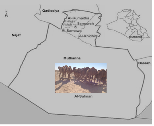
This study was performed on four camel herds in Badiat Al Samawah/Al Muthanna governorate 280 kilometers of southeast of Baghdad. It has dry desert weather. This area is sandy with ridges, and high populations of the Camelus dromedarius are living there. The area is covered with desert plants of diverse concentrations (Figure 1). The areas were surveyed during the period from February 2017 till February 2018. A total of 350 Camels were examined for the presence of hard ticks.
A- Ticks Collection
The ticks were collected during 15 visits to four camel herds in Bediat AL Samawah. On each occasion, all the visible ticks were collected from the body of each animal. Collected ticks were put in plastic vials with Isopropyl Alcohol for disinfection and fixing the samples. Ticks collected from each animal was put alone. Later on, ticks were transferred to the clinical pathology laboratory, College of veterinary
medicine/Al Muthanna university. After that the ticks were counted and then prepared for identification. Each tick was processed and identified microscopically according to the keys of hard ticks mentioned previously (Hoogstraal, 1956; Hoogstraal et al.,1981; Soulsby, 1986).
B- Preparation of Ticks Microscopic Slides
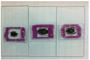
Specimens of ticks containing host tissue in their mouth are cleaned then placed in 10% KOH for softening, small specimens required 2 days in 35°C, while large specimens required 2 weeks in 35°C. The larva and nymph required one day only in 35°C. Later on, the samples were mounted on glass slide using Canada balsam and simple modified technique using a small rounded wall of clay to raise the cover slides from the mounted tick (Figure 2). The prepared samples were examined using simple inverted microscope. All information concerning number and genus of ticks was recorded during examination of each sample. The number of male, female and the immature stages of ticks were also recorded.
Table 1: Shows the overall and sex wise infestation in camel
| Criteria | No. and the percentages of female and male | No. of infested animal | Percentages of infestation % to the total number of the specific species (female or male) | Percentages of infestation % to the total number of examined camels | Percentages of infestation % to the total number of the specific species (female or male) |
| Female | 330 (94.28%) | 325 | 98.48 % (325 out of 330) | 92.85% (325 out of 350) | 98.48 % ( 325 out of 330) |
| Male | 20 (5.71%) | 15 | 75 % ( 15 out of 20) | 4.28% (15 out of 350) | 75 % ( 15 out of 20) |
| Total number | 350 | 340 | 97.14 % |
Results
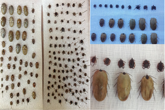
A total, 1455 ticks were collected during the study period (Figure 3).
The overall prevalence of the tick’s infestation was 97.1% (340 out of 350). The highest infestation rate was 98.48 % (325/ 330) in female, while, the male percentage was 75% (15/20). Meanwhile, the ticks infestation percentages out of the total number (350) examined camels were 92.85% (325 out of 350) and 4.28% (15 out of 350) for female and male respectively (Table 1).
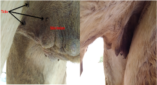
The skin lesions appeared on the ticks infested camels were classified as mild, moderate and severe lesions depending on the degree of the damage in the infested area. Moreover, the majority of the examined animals expressed severe lesions particularly on the udder (Figure 4).
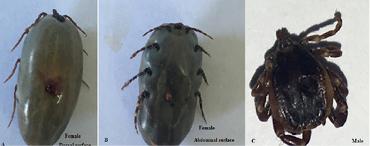
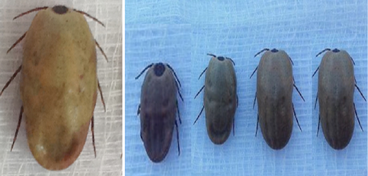
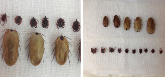
Nevertheless, in regard to the number of infested ticks per each individual camel, the severity of ticks infestation were categorized as low, medium and high with 15-60, 61- 200 and above 201 ticks per animal respectively (Table 2). The percentages of mild, medium and high infestation were 22.94%, 27,94%, and 49.11% respectively. The ticks were found on different site on the body (legs, chest, axillary, udder, testes, anus, inguinal, ear and face). The heavy infestation was found beneath the tail, udder, chest and inguinal area. The most abundant species of ticks found on the camels at these study areas were Hylomma dromedarii (Figure 5) and Boophilus spp. (Figure 6).Ticks were found as larvae, nymph and adult stages (Figure 7).
Table 2: Shows the classification of the severity of tick infestation.
| Criteria | Number of animal | Percentage % |
| Low (15-60 ticks per animal) | 78 | 22.94 % |
| Medium (61- 200 ticks per animal) | 95 | 27.94 % |
| High (above 201 ticks per animal) | 167 | 49.11% |
| Total number of infested animals | 340 | 100% |
Discussion
Ticks are the supreme important between the factors distressing camels health in spreading different diseases causing agents and casue blood loss, damage to hide and udder (Teka et al., 2017; Al Salihi et al., 2017; Walker et al., 2003; Higgins, 1983). Ticks are caused a significant economic loss in a camel productivity (Zeleke and Bekele, 2004). The results of the present study revealed that the Camelus dromedarius in Badiat Al Samawah harbour considerable level of numerous species of ticks. The occurrence of ticks in the present study has been likewise reported by different authors in another governorates in Iraq, albeit there is variation in the level of infestation in such reports (Hussein and Al-Fatlawi, 2009; Mohammad, 2015; Shubber, 2014: Shubber et al., 2014). The results of the present study revealed very high infestation rates between examined camels with an overall infestation percentage 97.14 % (340 out of 350). This result is compatible with previous reports (Mohammad, 2015; Hussein and Al-Fatlawi, 2009), meanwhile, it is disagreed with Shubber et al., (2014), who reported lower infestation percentage (65.77 %) in camels. The totally high infestation percentage (97.14%) reported in the present study pointing that the camel obtains extra parasitic burden of ticks because the lifestyle practiced by its owners. It is worthwhile to mention that Bedouins rear large number of camels and looking on various ecosystems of the study area including border of the cities, villages, semi-desert and desert enabling the camels collect high number of ecto-parasites especially various ticks species. Furthermore, the high ticks infestation percentage reported in the present study are in agreement with previous percentages reported elsewhere in the world as, 85.5%, 78.6 % and 83% in Iran (Champour et al., 2013), Eastern Ethiopia (Teka et al., 2017) and Pakistan (Javaid et al., 2013) respectively. However, this result is incompatible with Abdurahman, (2006), who reported 49.1% tick infestation on the camels because the examination of the udder region only. Moreover, it is also disagreed with Hegazy et al. (2004), who reported ticks infestation on eyelids of 12 out of 488 examined camels in Egypt, and this low infection probably due to the examination of eyelids only.
The result of the present study revealed that the highest infestation rate was 98.48 % (325/ 330) in female, while, the male percentage was 75% (15/20). This result is compatible with previous researcher who revealed heavy infestation of ticks in female (Yakhchali, 2006; Lees and Miline, 1951). The high reported infestation percentage in female might be due to the large number of the examined female (330) in compare to male (20) as always there is low proporation of the male rear to the female in the camel herds because of its ferocity. Two genus of hard ticks (Hylomma dromedarii and Boophilus spp) were determined in the present study and this result is in agreement with Hussein and AL- Fatlawi, (2009) who also reported the same two species in Al-Qadisiya province. These results are also endorsed by the previous studies elsewhere in the world (Javaid et al., 2013; Kady, 1998), who mentioned that Hylomma dromedarii was the common species infested camels. Some species of ticks had been isolated including Hyalomma spp. Amblyomma and Ripicephalus from Camelus dromedarius in Africa and Asia (Mukasa-Mugerwa, 1981). Addionally, Begum et al., (1970) invesitaged Hyalomma dromedaries and Javaid et al., (2013) found Hyalomma dromedarii, H.an.excavatum, H.impeltatum, H. an.anatolicum, H. marginatum and H. schulzei in Pakistan. Furthermore, Hyalomma, Boophilus and Ripicephalus spp.was invesitaged in Iran (Yakhchali, 2006). Howerver, some factors including variation of the climate, seasons and age of the animal are palying important role in the presence of the species of the tick.
The results of this study showed that the ticks were found on different site on the body (legs, chest, axillary, udder, testes, anus, inguinal, ear and face). The heavy infestation was found beneath the tail, udder, chest and inguinal area. This result is in agreement with the results reported previously in Iraq (Hussein and Al- Fatlawi, 2009) and Iran (Yakhchali, 2006). The distribution of the ticks overall parts of the body lead to develop skin lesion and mastitis, if severe infestation occurred on the udder (Al Salihi et al., 2017). The neglected treatment of ticks infestation is contributing in the continuity of the ticks irritation and their life cycle that lead to heavy tick burden infestation for the animals.
The results of the current study also reported the number of infested ticks per each camel , and the tick infestation burden percentages were 22.94%, 27.94% and 49.11% for mild, medium and high respectively. This results are compatible with results reported previously by Van and Jongejan (2000) and Javaid et al., (2013), who found a relatively heavy mean tick burden with very braod range in the number of ticks per camel (6-173 tick).
In conclusion, this study approved the heavy ticks infestation between the camels populations in Al Muthanna governorate. Consequently, ticks infestation lead to develop variation in severity of the clinical signs. The authors recommend another future studies that contribute to the understanding the role of ticks infestation in the transmission of serious diseases in camelids in Iraq. Moreover, education and awareness of the farmer and owner should be essential about the tick borne diseases and threats produce by the ticks to the animals. Planning of ticks eradication programs, facilities and drugs should be provide by the governmental veterinary authorities to control spreading ticks between livestockes.
Acknowledgements
The authors would like to thanks Camel’s owners for their hospitality, cooperation, patience and helping during collection of samples.
Conflict of interest
There is no conflict of interest for any authorities and the project was self-supported financially.
Authors Contribution
The authors are contributed equally, but the concept of the research was suggested by Assistant professor Dr. Karima Al Salihi.
References


