Journal of Animal Health and Production
Research Article
Nematode Infection Depresses Humoral Immune Responses of Goats to Mycoplasma capricolum subsp. capripneumoniae Vaccine Antigens
Maritim Kipkoech Noah1, Ngeiywa Moses Mwajar2, Siamba Donald Namasaka3, Mining Simeon Kipkoech4, Wesonga Hezron Okwaka5
1Department of Animal Sciences, Pwani University; 2Department of Biological Sciences, University of Eldoret;3Department of Agriculture and Veterinary Sciences, Kibabii University;4Department of immunology, Moi University; 5Veterinary Sciences and Research Institute, KARLO.
Abstract | Mycoplasma capricolum subsp. capri pneumoniae (Mccp) causes contagious caprine pleuropneumonia (CCPP), a disease of goats characterised with a high morbidity and mortality in non vaccinated goats. The objective of this study was to investigate the impact of nematode infection on humouralimmune responses to Mccp vaccine in goats. Forty (40) goats, aged 9 – 12 months, were randomly allocated to four groups of ten. Group A were orally inoculated with infective stages of nematodes followed by immunization with inactivated Mccp vaccine after 3 weeks. Group B were not inoculated with nematodes but immunized as Group A. Group C were inoculated with infective stages of nematodes but not vaccinated as in A. Group D was neither inoculated nor vaccinated. Clinical observations and records were done daily at 8.30 AM, blood for sera analysis was collected weekly, while pathological data was collected at post-mortem. Analysis of variance and Tukey Honest Significant Difference, a post hoc test, multiple comparisons of means were performed. The results showed that immune response to Mycoplasma vaccine antigens in helminth infected group (A) was significantly lower than that in vaccinated none helminth infected (B) group (p<0.05).Evidence from this study indicates that worm infection impacts negatively on immune response to vaccine antigens. Thus, we recommend that deworming exercise should be carried out before planned vaccinations are carried out.
Keywords | Contagious caprine pleuropneumonia, Nematodes, Goats, Vaccination, Humoral immunity
Editor | Asghar Ali Kamboh, Sindh Agriculture University, Tandojam, Pakistan.
Received | April 15, 2018; Accepted | May 05, 2018; Published | June 25, 2018
*Correspondence | Maritim Kipkoech Noah, Department of Animal Sciences, Pwani University; Email: [email protected]
Citation | Noah MK, Mwajar NM, Namasaka SD, Kipkoech MS, Okwaka WH (2018). Nematode infection depresses humoral immune responses of goats to mycoplasma capricolum subsp. capripneumoniae vaccine antigens. J. Anim. Health Prod. 6(2): 57-61.
DOI | http://dx.doi.org/10.17582/journal.jahp/2018/6.2.57.61
ISSN (Online) | 2308-2801; ISSN (Print) | 2309-3331
Copyright © 2018 Maritim et al. This is an open access article distributed under the Creative Commons Attribution License, which permits unrestricted use, distribution, and reproduction in any medium, provided the original work is properly cited.
Introduction
Mycoplasma capricolum subsp. capri pneumoniae (Mccp), a member of the order Mycoplasmatales and class Mollicutes (OIE, 2014; MacVey et al., 2013), causes contagious caprine pleuropneumonia (CCPP) a highly contagious bacterial disease characterized by high morbidity of up to 100% and mortality of up to 80%. Also characterized by respiratory distress, mucopurulent nasal discharge, fibrinous pleuropneumonia afflicting mainly goats (OIE, 2017) more so in non- vaccinated flocks. Transmission occurs predominantly through direct contact mediated by aerosolization of the respiratory secretions to a greater extent when there is overcrowding as at water points and long distance spread through wind under optimal conditions has also been cited (MacVey et al., 2013). Immunosuppression due to nematode infection is reflected in a dampened resistance to concurrent infections such as buffalos’ susceptibility to bovine mycobacterium infection (Ezenwa and Jolles, 2011) and poor response to vaccine antigen reflected by low seroconversion (Urban et al., 2007) or none at all. There is evidence that ruminant helminths are capable of modulating host immune response (Anthony et al., 2007; Shin et al., 2009; McNeilly and Nisbet, 2014).
This highly contagious disease (CCPP) was first described in Algeria in 1873 and in South Africa in 1881 (OIE, 2014). The aetiological agent, Mccp, was first isolated and identified in Kenya (OIE, 2014).The disease in goats has been reported in several Eastern African, Asian and Eastern European countries (El-Deeb et al., 2017; Nicholas and Churchward, 2012; Awan et al., 2010; Srivastava et al., 2010; Centikaya et al., 2009; Ozdemir et al., 2005).
A significant small ruminant population in Kenya is found in the arid and semi-arid lands (MoLFD, 2009) that border National game parks /reserves teeming with wild ruminants. CCPP was also confirmed in captive wild ruminants kept in a wildlife reserve in Qatar where it involved wild goat (Capra aegagrus), nubian Ibex (Capra ibex nubiana), Laristanmouflon (Ovisorientalis laristanica), and Gerenuk (Litocranius walleri) (Arif et al., 2007). Mccp was isolated in 15.4% of samples submitted from sheep in East Turkey (Centikaya et al., 2009). The isolation of Mccp in sheep and wild ruminants’ samples is of particular importance to Kenya since a significant sheep and goat population is found in the arid and semi-arid lands that include or border the National game parks and wildlife reserves teeming with game small ruminants. The pasture in regions where the disease is endemic is sometimes heavily contaminated with helminth infective stages during rainy seasons leading to high infestation of the goats and other livestock. Vaccination of goats using an inactivated Mccp antigen suspended in saponin is protective for up to 14 months is usually done to protect goats from the clinical disease in Kenya however; revaccination is recommended annually (Rurangirwa et al., 1987, OIE, 2014). The objective of this study was to investigate the effect of nematode infection on humouralimmune response in goats to killed Mycoplasma vaccine antigens.
MATERIALS AND METHODS
Ethical Statement
This research was approved by the Institutional Animal Care and Use Committee of KARLO-Veterinary Science Research Institute, Muguga North upon compliance with all provisions vetted under and coded: KALRO-VSRI/ IACUC012/22092017.
The experimental study was carried out in goats housed in pens in laboratories under microbiology and parasitology divisions of veterinary sciences research institute, KARLO Muguga North where nematode larvae used in this experimental infection were also cultured.
Experimental Design
Forty, dewormed with Albendazole (ValbazenR,Norvatis) at 10mg/Kg body, CCPP free and unvaccinated goats, aged between 9-12 months, were used in the study. The goats were randomly allocated to four groups, (A,B,C,D) of 10 animals each. Helminth status was confirmed after the observatory period of 28 days.
Goats in group A were inoculated orally with 3000 mixed cultured larvae (L3) suspended in 10ml of water (300 L3/ml) then flushed down with 10 ml of phosphate buffered saline. Three weeks after inoculation with infective larvae (49th day of study) the group was vaccinated with 1 ml of inactivated Mycoplasma capricolum subsp. capri pneumoniae vaccine (CaprivaxR) from Kenya Veterinary Vaccine Production Institute (KEVEVAPI).
Animals in group B (n=10) with no detectable worm infection were given 10 ml of normal saline without helminths. Vaccination with Mycoplasma antigen was done on the 49th day of study as described for group A. Animals in group C (n=10) were inoculated infective larval stages as described for group A but not given Mccp vaccine. Group D goats were given 10 ml of phosphate buffered saline orally but were neither inoculated with worms nor vaccinated with Mycoplasma antigen.
All the goats in the four groups were bled for sera on weekly basis. Faecal samples for egg per counts were taken per rectal every fourteen days. The sera were frozen to be used later to monitor seroconversion in response to the vaccine using cELISA.
Blood for cELISA
Blood samples were investigated for serum late antibodies to M. capricolum subsp. capri pneumoniae antigen on vaccination by a modification of a competitive ELISA (c-ELISA) used as described by Wesonga et al. (2004).
Parasitological Observations
Modified McMaster Technique (Zajac and Conboy, 2012) was used on faecal specimens collected directly from the rectum of the experimental goats for faecal egg counts.
Statistical Analysis
All data analyses were performed using R statistical package (Rx64 3.2.4 Revised) and compared with Minitab® statistical software output. Analysis of variance was performed to ascertain the difference among and between the group means of antibody response to Mycoplasma vaccine antigen in goats with or without helminths. P values less than 0.05 were statistically significant. To establish which of the groups differed from the others, a Post Hoc Test, Tukey honest significant difference (HSD) multiple comparisons of means giving 95% family-wise confidence level wasperformedon all data sets.
RESULTS
Faecal Egg Per Gram Counts in Worm and Non –Infected Goats
The faecal egg count in groups A (Table 1) increased with time from the initial count of non detectable egg per gram to the 8th week when the trial was terminated. Both groups
Table 1: Summary of mean egg per gram counts in worm and non–infected goats over a period of eight weeks.
| Week 2 | Week 4 | Week 6 | Week 8 | |||||||||||||
| Group | A | B | C | D | A | B | C | D | A | B | C | D | A | B | C | D |
| Mean |
206. 250a |
11. 111b |
200. 000a |
6.25 b |
718. 750a |
105. 556c |
381. 250b |
75. 000c |
1500. 0a |
0.0b |
1212. 5a |
-0. 0b |
3775a |
22. 22b |
3475. 00a |
43. 75b |
| STDV |
129. 3873 |
22.0 4793 |
136. 277 |
17.6 7767 |
303. 4769 |
39. 0868 |
194. 4544 |
46. 291 |
151. 1858 |
0 |
419. 8214 |
0 |
517. 5492 |
36.3 2416 |
2141. 261 |
41.7 2615 |
| SED |
45.7 4532 |
7.7 9512 |
48.1 8121 |
6.25 |
107. 2953 |
13.8 1927 |
68.75 |
16.3 6634 |
53.4 5225 |
0 |
148. 4293 |
0 |
182. 9813 |
12.8 4253 |
757.4 0502 |
14.7 5242 |
STDV Standard deviation; SED Standard error of a difference
showed a similar growth pattern in faecal egg count as the worms matured. The mean egg per gram for group A and C increased from non detectable in the first week to a mean of 3775 and 3475 at the end of week eight when trial period ended.
Group B and D (Table 1) started with non-detectable faecal egg count; some goat droppings had detectable helminths eggs at the end of the 4th week with the highest count being 150 eggs per gram of faeces. The goats were then dewormed to reduce worm infection to the lowest levels. One-way analysis of means output showed a statistically significant difference between the group means. p value 7.098e-08which is less than 0.05. A Tukey post hoc pair wise comparison was performed with the output showing that the difference in means is significantly different between the groups B-A (p-value=0.0000021), D-A (p-value=0.0000039), C-B (p-value=0.0000755) and D-C (p-value=0.0001211) only.
Immuneresponse to Killed or Inactivated Mycoplasma capricolum subsp. Capri pneumoniae Vaccine Antigen
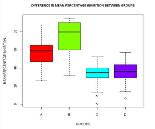
Goat serum samples with percentage of inhibition greater than or equal to 50% were considered positive for presence of Mycoplasma capricolum subsp. capripneumoniae antibodies. Figure 1 shows a box plot depicting variation in antibody response reflected by percentage of inhibition means such that for group A titres increased steadily to peak on the 7th week at 64.79 percentage inhibition (PI) while for group B it increased steadily to peak at the 4th week PI=85.97 without other appreciable increase until termination of study. Group C and D goats had PIs that did not at any time surpass the 50% PI mark which is a non significant antibody immune response in this kind of test.
Visually the box plot (Figure 1) shows that some means were different. One-way analyses of means showed a significant difference between the groups with and without nematode infection (p-value equal to <2e-16 which is less than 0.05).
Tukey multiple comparisons of means giving 95% family-wise confidence level was conducted and a statistically significant difference between groups A and B, A and C, A and D, B and C, B and D (p-values = 2.7e-10, 1.2e-15, 2.9e-13, < 2e-16, and < 2e-16, respectively) was observed.
DISCUSSION
Antibody response reflected by percentage of inhibition means for group A (vaccinated when nematode infected) peaked on the 7th week at 64.79 PI while group B (vaccinated without detectable nematode eggs) peaked at the 4th week PI=85.97. In groups C and D (not vaccinated) PIs did not at any given time surpass the 50% PI mark which is a non significant antibody immune response in this kind of test.
From one way analysis of variance there was a significant difference between vaccinated groups with or without nematode infection and those that were not vaccinated (p-value equal to <2e-16) which is less than 0.05.
A Post Hoc Test compared groups A (vaccinated with worms) with B (vaccinated without worms) showed a statistically significant difference (p-values = 2.7e-10) suggesting that there was a strong nematode influence on antibody response. This compares with reports from investigators using other models who have shown that helminths can influence vaccine efficacy by modulating host immune responses in particular when Th1- like and cellular – dependent responses are required (Stelekati and Wherry, 2012; Stelekati et al., 2014). The difference in antibody response in worm infected and none infected groups also concurs with the findings of Elias et al. (2005) and Van Riet et al. (2007) that Schistosoma sp. and Onchocerca volvulus infection decreases efficacy of vaccine against tuberculosis and tetanus respectively. Urban Jr. et al. (2007) and Steenhardet al. (2009) were also able to show, in separate investigations, that Ascaris suum co-infection alters seroconversion and efficacy of vaccine against Mycoplasma hyopneumoniae. Working with mice, Su et al. (2006) reported that H. polygyrus was able to down regulate the strong immunity against Plasmodium chabbaudi induced by blood-stage antigens. On further analysis, groups C-A, D-A, C-B and D-B were compared; a statistically significant difference was observed for the paired groups. But there was no statistical significant difference between group D (none vaccinated, none helminths infected) and C (helminths infected none vaccinated). This clearly suggests that the difference in antibody response was due to vaccine antigens as indicated by percentage inhibition less than the cut off point for groups C and D. The lower antibody response in group A compared with that of group B also suggests that the internal parasites may have had an impact on immune responses to bystander antigens such as those from infective agents and vaccines. This is in agreement with reports by Salgame et al. (2013) and Chatterjee et al. (2015) that helminths have an influence on immune response to disease and vaccine antigens.
Conclusions
From the above discussion, there is evidence indicating that worm infection has a negative influence on immune response to bacterial infection and bystander antigen such as those from vaccines. Immune response to inactivated Mycoplasma vaccine antigens in helminths infected groups was lower than that in none helminth infected group (p value=2.7e-10). None helminth infected group had higher means suggesting that helminths infection affects immune response to vaccine antigens.
To further characterize the effect of helminths infections on the host response to microbial pathogens and vaccines more extensive randomized studies with the use of relevant measurable biological indicators such as specific cytokines to assess actual immune responses are required. However, animal welfare concerns and the cost involved restrict animal use in such experimental studies. This complex response includes both type II immune response as well as innate and adaptive immunoregulatory compartments. We therefore recommend that livestock be dewormed before vaccination is carried out.
ACKNOWLEDGEMENTS
To the Director of Veterinary Sciences Research Institute (VSRI), Kenya Agricultural & Livestock Research Organization (KALRO), Muguga North for accepting me to work in the institution.
To National Commission for Science and Innovation for the support they gave towards completion of this work.
To Professor P. P. Semenye, NUFFIC-NICHE KEN 212 for the support the project accorded me toward completion this study.
Conflict of interest
There is no conflict of interest.
Authors Contribution
Moses Ngeiywa: Project supervisor and course work instructor.
Donald Siamba and Simeon Mining: Supervisor.
Hezron Wesonga: Gave guidance during experimental inoculation in goats.
REFERENCES


