Research Journal for Veterinary Practitioners
Case Report
Iatrogenic Uterine Perforation Resulting in Fetal Maceration in an Ewe
Garba Bashiru1*, Abubakar Nura2, Bodinga Hassan Abubakar2, Oviawe Ime Ekaete2, Ahmad Umar Salisu2, Garba Usman Rambo3
1Department of Veterinary Public Health and Preventive Medicine, Faculty of Veterinary Medicine, Usmanu Danfodiyo University, Sokoto, Nigeria; 2Department of Veterinary Surgery and Radiology, Faculty of Veterinary Medicine, Usmanu Danfodiyo University, Sokoto. Nigeria; 3Veterinary Teaching Hospital, Faculty of Veterinary Medicine, Usmanu Danfodiyo University, Sokoto. Nigeria. PMB 2346, City Campus, Sokoto. Nigeria.
Abstract | Fetal maceration is the disintegration of a fetus that can occur at any stage of gestation. Maceration is common in almost all the animals; however; it is rare in case of the sheep. Maceration is common cause of mummification and generally occurs in the event of the death of a fetus after the formation of the fetal bones beyond 100 days in small ruminants, such animals failed to abort, although the cervix is almost open. Complications often arise after traditional medical intervention, especially in obstetrical cases. Iatrogenic uterine perforation can present an adverse effect, including abortion or fetal death and mummification. The present case report is a rare case of uterine perforation due to a failed attempt to manage a swelling noticed on the ventral abdomen. Examination of the ewe revealed the presence of opening on the ventral abdomen with purulent discharge and macerated fetus by the presence of dismembered bones within the uterus. After a radiograph revealed the presence of disintegrated fetus, an emergency laparotomy was performed to evacuate the autolysed fetus and the flushing of the uterus. Subsequently, antibiotic treatment to clear the remnant of the pyogenic bacteria.
Keywords | Fetal maceration, Uterine perforation, Radiological investigation, Ewe, Laparotomy
Received | February 01, 2020; Accepted | April 03, 2020; Published | May 28, 2020
*Correspondence | Garba Bashiru, Department of Veterinary Public Health and Preventive Medicine, Faculty of Veterinary Medicine, Usmanu Danfodiyo University, Sokoto, Nigeria; Email: [email protected]
Citation | Bashiru G, Nura A, Abubakar BH, Ekaete OI, Salisu AU, Rambo GU (2020). Iatrogenic uterine perforation resulting in fetal maceration in an ewe. Res J. Vet. Pract. 8(2): 15-19.
DOI | http://dx.doi.org/10.17582/journal.rjvp/2020/8.2.15.19
ISSN | 2308-2798
Copyright © 2020 Sayeed et al. This is an open access article distributed under the Creative Commons Attribution License, which permits unrestricted use, distribution, and reproduction in any medium, provided the original work is properly cited.
Introduction
Traditional and alternative medicine which is a phrase used to describe a set of healthcare practices delivered outside the conventional healthcare system is still popular among indigenous Africans (James et al., 2018). In Nigeria, traditional medical interventions encompass administration of local herbs, as well as indigenous healthcare practices including bone settings, and cauterisation. The practice is widespread, and a considerable number of Nigerians rely on such practices to heal and maintain their health (Kasilo and Trapsida, 2010). One of the contributing factors driving patronage is poverty and lack of accessibility to new biomedical services, particularly among remote locations (Krah and Ragno, 2018). However, despite its perceived benefits, traditional remedies are associated with lots of risks and undesirable effects (Krah and Ragno, 2018).
Iatrogenic illnesses are those adverse health conditions that follow diagnostic and therapeutic procedures undertaken on a patient or other forms of medical interventions (Chauhan et al., 2013). Although more commonly associated with modern medical interventions, including adverse drug effects, its original meaning may extend to health conditions that result from remedies offered by traditional healers. One of such practices in livestock in Nigeria is the use of a hot knife or metal spear to do branding, cauterisation or lancing of infected skin lump, e.g. boil. Customarily, indication for such intervention is the presence of swelling (bulge) on any part of the integument that persisted for several days. The procedure entails, heating the metal object until red hot, and placing it on the affected part followed by application of pressure to lance and supposedly drain the lump. When conducted by experienced healers, the results are usually successful. However, complications can arise when the procedure is not done correctly, including perforation or third-degree burn (Irvin et al., 2003).
Case report
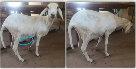
A one and half year-old Balami-Cross ewe was presented to the Veterinary Teaching Hospital of the Faculty of Veterinary Medicine, Usmanu Danfodiyo University, Sokoto, Nigeria with the complaint of swelling noticed at the ventral abdomen near the udder (Figure 1). The client claimed to have noticed the swelling two weeks before presentation.
Clinical examination
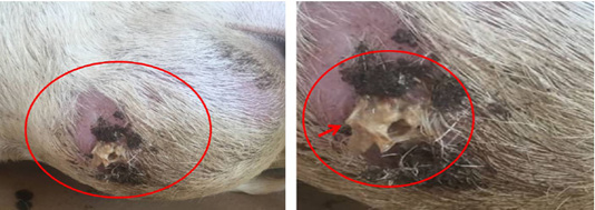
The ewe was observed to be alert and active. The rectal temperature was 39.4 oC, pulse and respiratory rates were 83 beats and 100 cycles per minute, respectively. The animal was restrained and placed on left lateral recumbency in order to perform a detailed examination. A reddish-grey foul-smelling discharge was noticed from the burst swelling with protruding bony structures (Figure 2). On examination of the external genitalia, the cervix was found closed with no discharge.

Figure 2: The patient on presentation showing the burst swelling on left lateral recombency. The circumscribed area shows the pus with protruding fragment of fetal bone (arrow head).
Laboratory and radiological investigations
Blood samples were collected via jugular venipuncture for haematological analysis (Table 1). Similarly, faecal samples (per rectum) and swab sample from the pus discharge were also collected for routine parasitological and microbiological isolation and sensitivity test.
Parasitological investigation revealed the presence of Eimeria oocyst (+) and scanty Strongyle eggs. Bacteriological isolation, on the other hand, indicated the presence of Eschericia coli and Salmonella species, while the antibiotic sensitivity test revealed that the Salmonella isolate is susceptible to streptomycin and gentamycin, respectively.
Table 1: Haemtological profile of patient.
| Parameters | Value |
| PCV (%) | 28 |
| Hb (g/dl) | 9.0 |
|
RBC (cells/mm3) |
9.78 × 10⁶ |
| Differential leukocyte count (%) | |
| Neutrophils | 45 |
| Lymphocytes | 55 |
| Monocytes | 0 |
| Basophils | 0 |
| Band cells | 0 |
Furthermore, lateral radiograph of the abdominal region of the ewe revealed evidence of fetal bone fragments within the uterus (Figure 3). The radiograph also revealed the fetus in a state of autolysis within the uterus without being aborted. Hence, an exploratory laparotomy was recommended.

Figure 3: Lateral radiograph of the abdominal cavity showing the dismembered fetal bones. Red arrows showing dismembered fetal bones.
Surgical procedure and management
The patient was prepared aseptically for the surgery. The hair was clipped, Surgical site was scrubbed using 4% Chlorhexidine gluconate (Savlon®, Vervaadingdeur, Johnson and Johnson (pty) Ltd, London), followed by methylated spirit (Rico Pharmaceutical IND. (Nig.) LTD, and then povidone-iodine (APACCO Pharmaceutical Nig. LTD, Ogun state Nigeria). The local anaesthesia was achieved with 2% lignocaine (Debocaine® ALDebeiky pharmaceutical industries Co.) using a ring block technique.
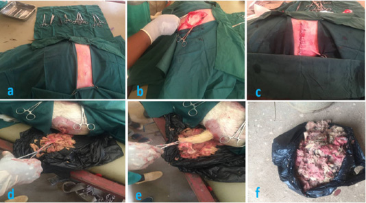
An incision was made to extend the opening at the ventral abdomen, and the fetal bones and viscera which weighed 2.6 kg were evacuated. The uterus was thoroughly lavaged using normal saline. After removing the macerated fetus, the uterus was adhered to the abdominal wall firmly. Adhesion was detached, and the uterus was closed with lambert suture pattern using Vicryl (Covenant root Nig. LTD, Shanghai SNWI Medical Co., LTD) size 0. The abdominal muscle layers were closed routinely with Vicryl size 1 using horizontal mattress suture pattern. Finally, the skin was sutured with nylon size 2 using ford interlocking suture pattern (Figure 4).

Figure 4: Showing the surgical procedure undertaken. (a) Triangular draping of the surgical field. (b) Incision into the abdomen via the paralumber fossa in order to exteriorize the punctured uterus for lavage and treatment. (c) Removal of the macerated fetus via the external opening. (e) Pulling of the skin remnant from the macerated fetus. (f) Complete macerated fetus removed from the uterus.
During the postoperative follow-up, 4 ml PenStrep® (De Santos pharm CO. LTD. Anambra State, Nigeria) was administered intramuscularly for five days against the possible remnants of the pyogenic bacteria as well as against secondary infection. Analgesia was achieved using a non-steroidal anti-inflammatory drug (Diclofenac Sodium; North China pharm. CO. LTD. Shijiazhuang, China) at the dose rate of 10 mg/kg for three days while multivitamins at 1 ml/kg were administered intramuscularly for five days to enhance healing and boost appetite.
Discussion
Fetal maceration is a destructive aseptic process that ensues when a dead fetus remains in-utero. It is also used to describe autolytic changes that usually follow antepartum death (Bozkurt et al., 2018). Upon intrauterine fetal death, maceration of the fetus commences by enzymatic autolysis of cells and generalized degeneration of connective tissues leading to brownish, tan, or yellowish skin discolouration and desquamation, overlapping cranial bones, loose joints, and formation of bullae which is preceded by skin peeling, as well as edema of the outer and inner organs with turbid effusions inside the fetal body and amniotic cavity (Bamber and Malcomson, 2015). Moreover, embryonic death and maceration are mostly associated with a variety of microorganisms inhabiting the uterus (Muin et al., 2018). Other means by which pathogenic bacteria can enter the uterus is through a dilated cervix or following rupture of the uterus leading to communication with the exterior of the abdominal cavity. When this happens, the soft tissues become digested by the combination of putrefaction and autolysis, leaving a mass of fetal bones in the uterus (Bozkurt et al., 2018).
According to the information received from the client, the ewe did not show any signs of ill health, nor was there any discharge from the vagina; except for the swelling observed at the ventral abdominal region. It is not uncommon for clients to claim ignorance when presenting patients to the hospital, especially if they perceive they are at fault in terms of any form of intervention instituted. Additionally, it is a common practice in Northern Nigeria for traditional healers to use hot metal to lance swellings on the body as a traditional remedy (Caja et al., 2004). In such situations, complications can arise where due to inadequate restraint or lack of experience, the puncture can extend beyond the integument affecting other internal organs, which can also create a portal of entry for anaerobic bacteria. Therefore, utmost care is desired in managing animal health so as to prevent animal suffering and dealth. Adequate care during animl husbandry practices is recommended as it not only prevent reproductive problems, but reduce exposure to other infectious diseases like mastitis (following traumatic injury to the teats), pneumonic mannheimiasis, and contagious ecthyma (Garba et al., 2019; Jesse et al., 2018a, b; Zamri-Saad et al., 1999).
Meanwhile, uterine perforation can result from several causes which can either be iatrogenic or spontaneous (Caspi et al., 1996). The typical conventional iatrogenic causes include; dilatation and curettage, hysteroscopy and others (Irvin et al., 2003). However, other causes associated with traditional medical complications are; liver and heart failure due to consumption of toxic substances as well as mechanical or surgical practices such as the present case. These interventions have been reported to account for over 250-500 deaths per 100,000 illegal procedures (James et al., 2018). Laboratory investigations revealed salmonella and E. coli as the two isolates, with the salmonella being susceptible to gentamycin and streptomycin. Hence, the initial medication administered using Penicillin and Streptomycin was sustained. Although the bacteria commonly associated with putrefaction and maceration are Staphylococcus aureus or streptococci, salmonella and E. coli are equally important members of the pyogenic bacteria species (Michael, 1998). Therefore, it has become paramount where possible to thoroughly investigate the causal microbes associated with cases of fetal maceration including the routine bacteriological culture and in some situations tissue culture screening for possible viral agent (Schwarz et al., 2019; Lefebvre, 2015; Bala et al., 2018). Viral isolation could be carried out on either embroynated chicken egg (Kumar et al., 2015; Bala et al., 2018; Azmi and Field, 1993; Balakrishnan et al., 2017). Similarly, the elevated levels of White blood cells (neutrophils and lymphocytes) are both indicative of bacterial infection, especially pyogenic bacteria (Herbinger et al., 2016; Rosales, 2018). This observation was further confirmed by the radiological finding of dismembered bones within the uterus, which is indicative of third category fetal maceration.
Conclusion
The case discussed herein highlights the importance of extreme care and precaution while administering medication or remedies to livestock, especially during late pregnancy. It also seeks to discourage the use of traditional methods in dealing with health-related challenges. As observed, uterine rupture is linked with an unpredictable event. It is associated with terrible maternal and fetal prognosis. It also indicates that traditional practice of healing using sharp metal objects should be considered a risk factor for uterine rupture, which may come with grave consequences.
Authors Contribution
AN, BHA and UAS conducted the surgical procedure. OIE run and interpreted the x-ray, BG and GUR drafted the manuscript. All authors criticized and edited the draft manuscript before final submission.
Conflict of interest
The authors have declared no conflict of interest.
REFERENCES


