Therapeutic Effect of Guava Fruit Extract on Cadmium Induced Toxicity in Developing Mus musculus
Therapeutic Effect of Guava Fruit Extract on Cadmium Induced Toxicity in Developing Mus musculus
Asmatullah1, Chaman Ara1,*, Shagufta Andleeb2, Maha Tahir1, Beenish Zahid1 and Madeeha Arshad1
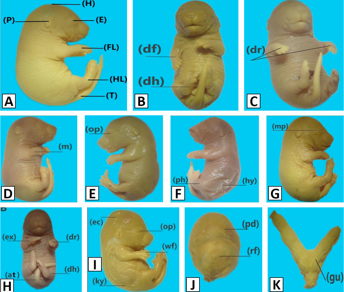
Macrophotographs of 18-day-old mouse fetuses recovered from mothers exposed to different doses of Cadmium on days 6 to 12 of gestation. A, from vehicle control; B, from antidote +6.25 μg/g BW; C, from antidote + 12.5 μg/g BW; D, from antidote+ 25.0μg/g BW. H, well developed head; E, well-formed eyes with closed eyelids; FL, well developed forelimbs; P, well developed pina; HL, well developed hindlimbs; T, well developed tail; op, open eyelids; dh, distorted hindlimbs; a, acheiria, m, meromelia, ec, exencephaly; gu, gravid uterus with resorbed fetuses; wt, wrinkled tail; m, micromelia, ph, phocomelia, ky, kyphosis; at, angular tail; hy, hygroma; rf, resorbed fetus; oe,openeyelid; dr, drooping wrist; wf, webbed feet; ec, excencephaly; pd, placental disc, ex, extended forlimb; df, distorted forelimbs; mp, microphthalmia.
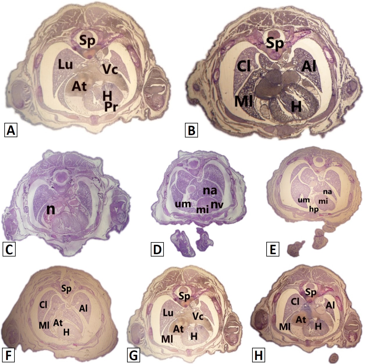
Microphotographs of transverse sections of 18-day-old mouse through liver region fetuses recovered from mothers exposed to different doses of Cadmium chloride and cadmium +antidote on days 6 to 12 of gestation. A, control; B, vehicle control; C, 6.25 μg/g B.W; D, from 12.5 μg/g BW; E, from 25.0 μg/g BW; F, from antidote +6.25 μg/g BW; G, from antidote + 12.5 μg/g BW; H, from antidote+ 25.0μg/g BW. Ml, middle lobe of right lung; Cl, caudal lobe; Al, accessory lobe; At, lumen of right atrium; H, heart; Sp, spinal cord; Vc, vena cava; Pr, pericardial cavity; um, underdeveloped middle lobe; nv, left ventricle is underdeveloped; mi, microcardia; na, necrosis in accessory lobe of lung; n, necrosis in atrium; hp, hypo pericardium.
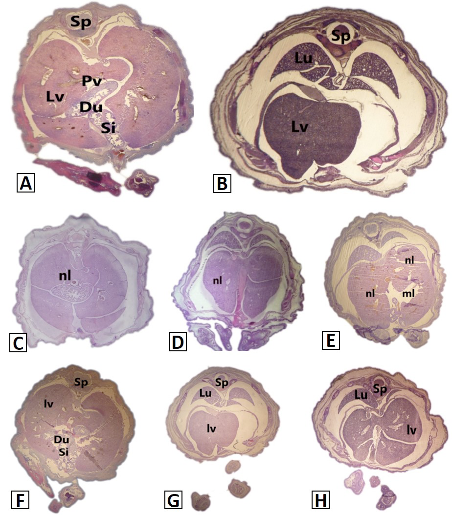
Microphotographs of transverse sections of 18-day-old mouse through liver region fetuses recovered from mothers exposed to different doses of Cadmium and cadmium +antidote on days 6 to 12 of gestation. A, control; B, vehicle control; C, 6.25 μg/g B.W; D, from 12.5 μg/g BW; E, from 25.0 μg/g BW; F, from antidote +6.25 μg/g BW; G, from antidote + 12.5 μg/g BW; H, from antidote+ 25.0μg/g BW. Sp, Spinal cord; Lu, lung; lv, liver; Du, duodenum; Si, loops of small intestine; ml, small portion of left lobe is missing; nl, necrosis.
A, control skeleton showing well ossified skeleton; B, from 6.25 μg/g BW; C, from 12.5 μg/g BW; D, from 25.0 μg/g BW. Cl, Clavical; Eo, Exoccipital; Fe, Femur; Fi, Fibula; F, Frontal; Ri, Ribs; U, Ulna; N, Nasal; Pm, Premaxilla; Z, Zygomatic; H, Humerus; So, Supraoccipital; Ip, Interparital; P, Parietal; R, Radius; Ro, Reduced ossification; Uo, Unossifie.
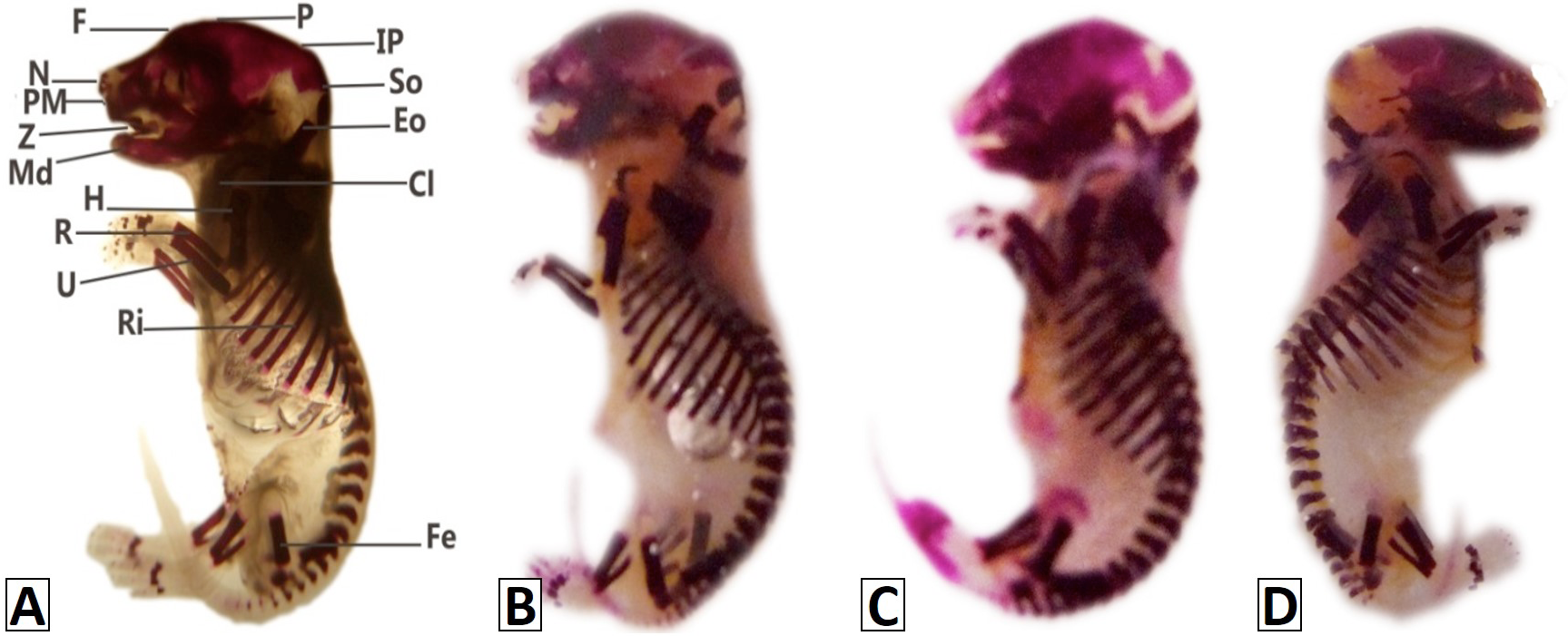
Macrophotographs of fetal skeletal preparations of 18-day-old mouse fetuses recovered from mothers exposed to different doses of Cadmium or antidote on days 6 to 12 of gestation. A, vehicle control; B, from antidote +6.25 μg/g BW; C, from antidote + 12.5 μg/g BW; D, from antidote+ 25.0μg/g BW. For abbreviations, see Figure 5.
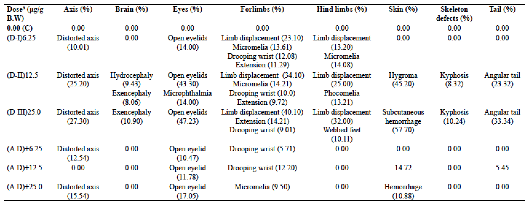
Developmental anomalies induced by cadmium in 18 day old mouse fetuses recovered from pregnant mice, administrated orally with different concentrations on days 6 to 12 of gestation.
Morphometric analysis of 18 day old mouse fetuses (Mean±SEM) recovered from pregnant mice, administered orally with different concentrations of cadmium (6.25, 12.5 and 25.0 μg/g B.W) and antidote +cadmium on days 6-12 of gestation.


