Taxonomic Divergence of Medically Important and Toxigenic Aspergillus minisclerotigenes from Aspergillus flavus
Taxonomic Divergence of Medically Important and Toxigenic Aspergillus minisclerotigenes from Aspergillus flavus
Amna Shoaib*, Zoia Arshad Awan and Naureen Akhtar
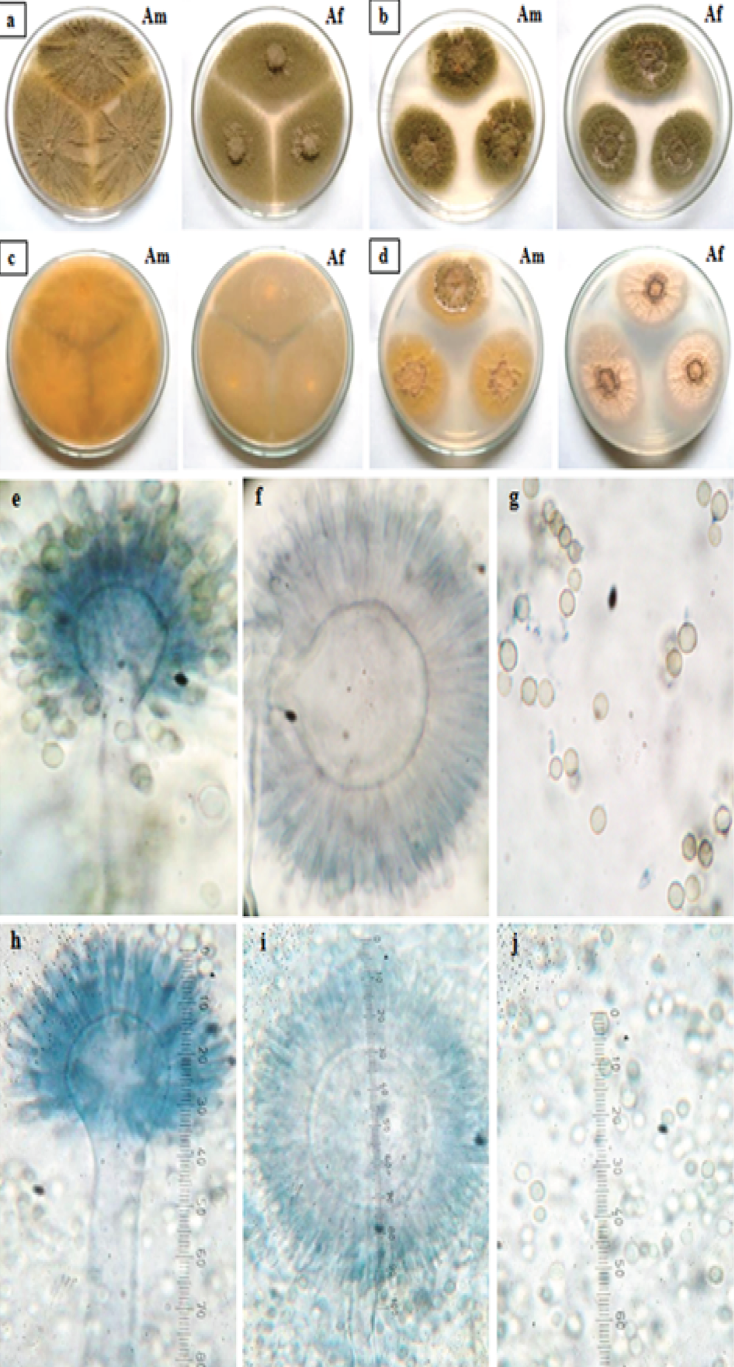
Comparison of colonies grown on MEA front and reverse (a and c) and on CZ (b and d). Microscopic study of A. minisclerotigenes (e-g) and A. flavus (h-j) showing seriation (uniseriate and biseriate) and conidial attachment. Am: A. minisclerotigenes; Af: A. flavus.
Comparative screening of aflatoxin production by A. minisclerotigenes and A. flavus grown on CCA. a: colony from front side; b: reverse colony; c: reverse colony under UV light. Am: A. minisclerotigenes; Af: A. flavus.
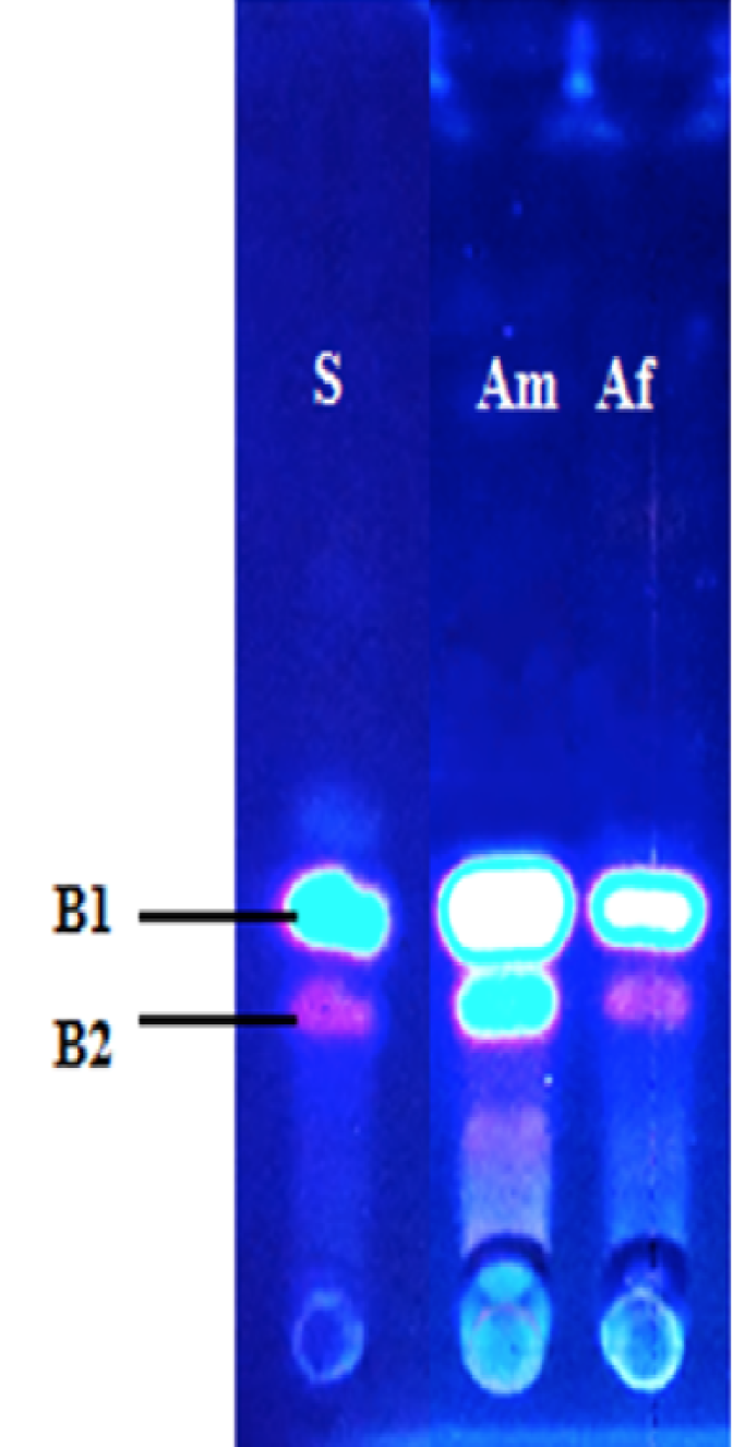
Aflatoxins production on TLC. S: AFBs Standard, Am: A. minisclerotigenes and Af: A. flavus.
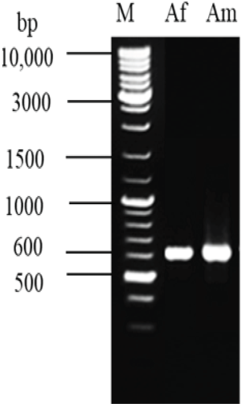
Amplified ITS region of strains, M=1kb DNA marker; Af: A. flavusus and Am: A. minisclerotigenes.
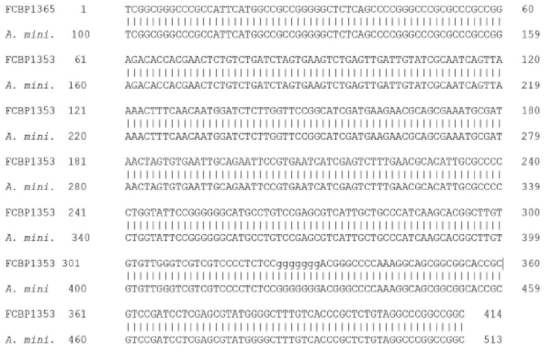
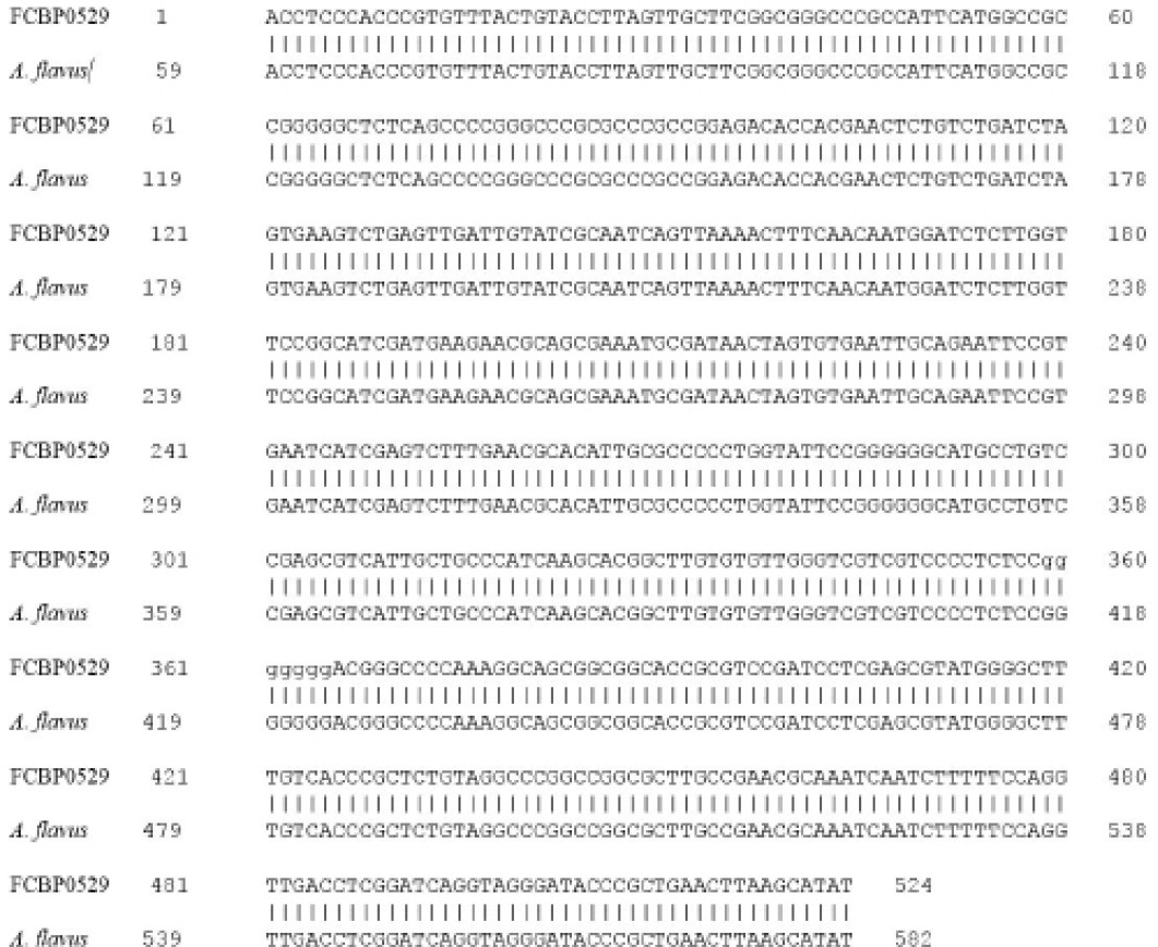
ITS sequence alignment of A. minisclerotigenes.
ITS sequence alignment of Aspergillus flavus.
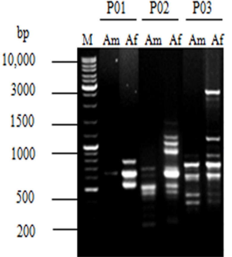
DNA banding profile of PCR-ISSR amplification product. M: DNA marker; Am: A. minisclerotigenes and Af: A. flavus.


