
Study the Effect of Allicin Nanoparticles In Thyroid Activity in Hyperthyroidism Experimental Induced Female Rats


Study the Effect of Allicin Nanoparticles In Thyroid Activity in Hyperthyroidism Experimental Induced Female Rats
Amal Mohammed Ali Alah Al-Marmadhi*, Saadeya Ali lefelef Al-Gnami
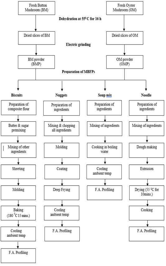
Product preparation flow chart of mushroom by products.1.
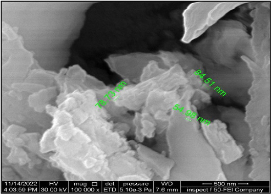
SEM of nanoparticles of appear elongated to irregular shape of allicin nanoparticles with particle size around 500 nm.
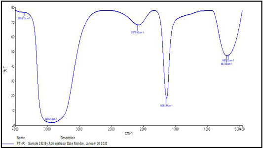
FTIR spectra.
A very broad peak.
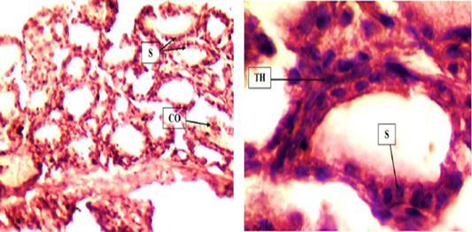
(A) Control group: Note normal thyroid follicles which lining with simple cuboidal epithelium (S) and these follicles contain few colloids (CO). 10X H & E. (B) Control group: Higher magnification, the thyroid follicles lined by single layer of simple cuboidal epithelium (S) and thin of interstitial tissue (TH). 40X H & E.
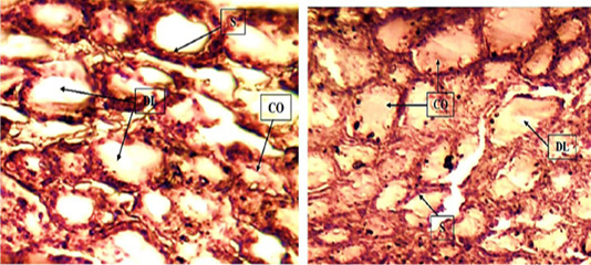
(A) T1 group: Thyroid tissue showed normal with dilated thyroid follicles (DL) and it contains colloid in their lumen (CO). Also these follicles line by simple cuboidal epithelium (S). (B) T1 group: There is dilation of thyroid follicles (DL) with large amount of colloid (CO), thin interstitial tissue and normal simple cuboidal epithelium which lined thyroid follicles (S). 10X H & E.
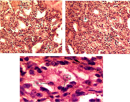
(A) T2 group: All thyroid follicles showed small in size and empty for colloid with hyperplasia of epithelial cells (H) which lined follicles, also there is high congestion of the blood vessels (C) and thickening of the interstitial tissue (T). 10X H & E. (B) Hyperplasia of epithelial cells which lining the follicles (H). The follicles showed small in size and devoid colloid (F). 10X H & E. (C) Higher magnification, the thyroid follicle showed hyperplasia (H) with desquamation (D) within the lumen of epithelial cells and thickening of interstitial tissue of thyroid. 40X H & E.

(A) T3 group: The thyroid follicles were dilated (DL) and contain large amount s of colloid (CO). Also there is thickening of the interstitial tissue of thyroid (T). 10X H & E. (B) Note high dilation of thyroid follicles (DL) with large amount of colloid (CO) and these follicles lined by simple cuboidal epithelium (S). 40X H & E.
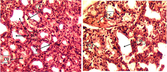
(A) T4 group. Some thyroid follicles showed dilation (DL) and lined by simple cuboidal epithelium (S) and it contains few colloid (CO). Other follicles were small in size with narrowed lumen and mild congestion of the blood vessels (C). 10X H & E. (B) Mild congestion of the blood vessels (C) and the thyroid follicles showed dilated (DL) and contain few colloids (CO). 10X H & E.

