
Sequence and Morphology of Cysticercus pisiformis in Local Breed Rabbits


Sequence and Morphology of Cysticercus pisiformis in Local Breed Rabbits
Kadhim Kh. K. Al-Khayat1*, Athmar K. A. Al-Azawi2
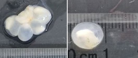
Shape and measurements of Cysticercus pisiformis cysts, grossly.
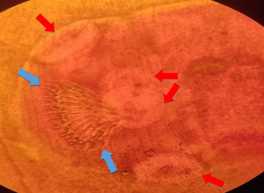
Structure of Cysticercus pisiformis Scolex staining by acetoacetic carmine stain, Suckers (red arrow) and Hooks (blue arrow), X40.
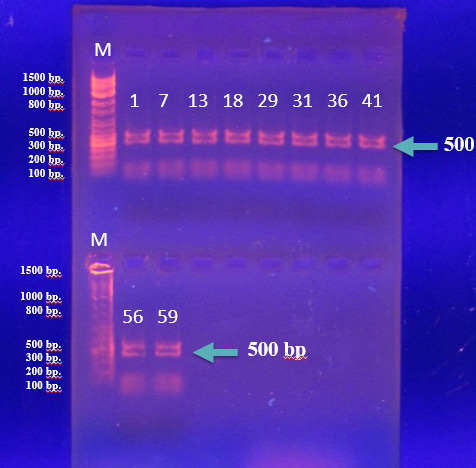
Agarose (1%) gel electrophoresis of PCR products for (NAD1) gene of C. pisiformis of rabbits and DNA Marker Ladder (M) (100- 1500bp), PCR products amplicons band lances (1, 7, 13, 18, 29, 31, 36, 41, 56 and 59) sized 500bp. (1h, 7 volt/cm2 TBE).
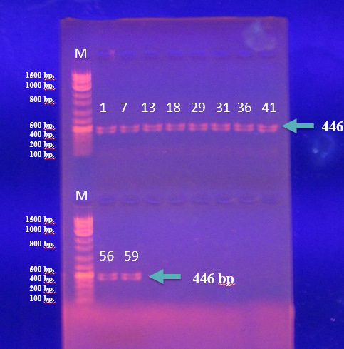
Agarose(1%) gel electrophoresis of PCR products for (COX1) gene of C. pisiformis of rabbits and DNA Marker ladder (M) (100- 1500bp), PCR products amplicons band lances (1, 7, 13, 18, 29, 31, 36, 41, 56 and 59) sized 446bp. (1h, 7 volt/cm2 TBE).

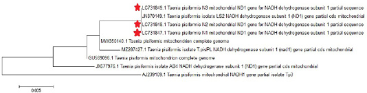
Phylogenetic tree study based on Taenia pisiformis NAD1 gene partial sequence.

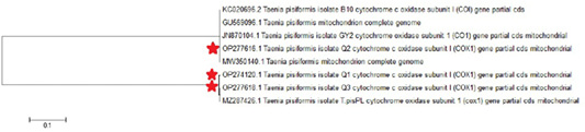
Phylogenetic tree study based on Taenia pisiformis COX1 gene partial sequence.

