Preliminary Evidence on Passage Attenuation of Orthoavulavirus javaense Genotype VII.1.1 and Its Potency as a Living Vaccine Candidate
Preliminary Evidence on Passage Attenuation of Orthoavulavirus javaense Genotype VII.1.1 and Its Potency as a Living Vaccine Candidate
Wahid Hussein El-Dabae*, Eman Shafeek Ibrahim, Eslam Gaber Sadek, Mai Mohamed Kandil
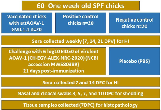
Diagram illustrating the details of in vivo experiment.
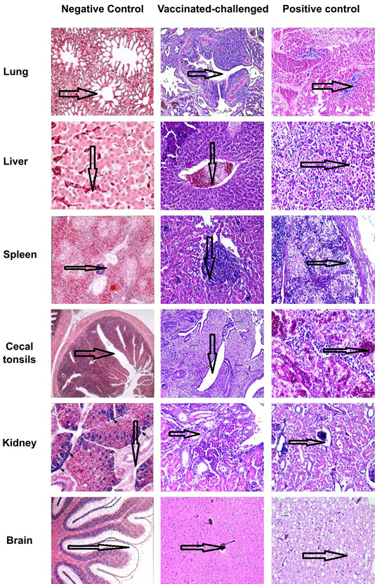
Histological examination of organs collected from different chick groups 7 DPC sections were stained with H & E stain with 20X magnification to identify different pathological lesions. Test group and positive control group showed a great difference in the severity of histological lesions compared with the normal histological appearance of the negative control group. The lung of the test group showed epithelial lining hyperplasia of secondary bronchioles, whereas the positive control group shows thrombus formation and blood vessel congestion. The livers of test group chicks showed thrombus formation, while the livers of the positive control group showed severe necrosis and sinusoidal thrombus formation. The spleen of test group chicks showed mild capillary sheath proliferation, while the spleen of the positive control group showed severe lymphocyte depletion and coagulative necrosis. The cecal tonsils of the test group showed lining epithelial necrosis and fibrinohemorrhagic exudates but those of the positive control group showed lining epithelial necrosis and fibrinohemorrhagic exudates. The kidneys showed congestion of blood vessels and vacuolar degeneration in both groups where milder lesions were evident in the test group. The brain showed demyelination and neuron degeneration with central chromatolysis which were milder in the test group.


