Pathological Study of Uterine Abnormalities in River Buffaloes (Bubalus bubalis) in Basrah
Pathological Study of Uterine Abnormalities in River Buffaloes (Bubalus bubalis) in Basrah
Batool S. Hamza1*, T.S. Al-Amery2
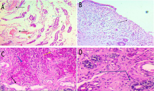
A: Acute endometritis: stromal edema (blue arrow) and severe engorgement of a small number of endometrial blood vessels (black arrows) H&E stain 100x. B: Acute endometritis: The endometrial epithelium showed localized denudation, as indicated by the black arrow. H&E stain 100x. C: Localized regions of subepithelial hemorrhage (blue arrow), together with congestion (yellow arrow) and edema (black arrow). H&E stain 100x. D: Acute endometritis: There is a moderate to mild infiltration of inflammatory cells, particularly polymorphonuclear and mononuclear cells, into the lamina propria (blue arrow). H&E stain 400x.
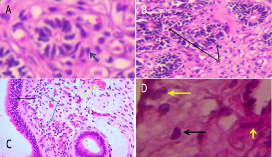
A: Acute endometritis: The glandular lamina and peri-endometrial glands show mild to moderate infiltration of mononuclear cells (blue arrow). 400X H&E stain. B: Acute endometris: enlargement of endometrial glands with degenerative changes (blue arrow) 100X H&E stain. C: Subacute endometritis: Marked hyperplasia of luminal epithelium (black arrow) with congestion (yellow arrow), edema and infiltration of inflammatory cells in lamina propria with glandular epithelial hyperplasia (blue arrow) (100x H & E). D: Subacute endometritis There was moderate to severe infiltration of mononuclear cells especially plasma cells (black arrow) as well as epithelioid cells in lamina propria around endometrial gland (yellow arrow) 400x H&E stain.
A: Subacute endometritis: The vascular medial coat thickened due to mild to moderate hypertrophy (black arrows), while the uterine glands atrophy (red arrows). 100x H & E stain. B: Chronic endometritis: Inflammatory cells infiltrating the lamina propria (green arrow) and low cuboidal endometrial epithelium (red arrow) (400x H & E). C: Chronic endometritis: the subepithelial zone has a heavy population of plasma cells and mononuclear cells (blue arrow). 400x, H&E stain. D: Chronic endometritis: Inflammatory cells infiltrate the periglandular fibrosis (red arrows). (400x H & E).
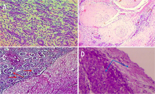
A: Chronic endometritis: uterine gland showing Atrophy of endometrial glands with infiltration of inflammatory cell in lamina propria (400x H & E). B: Chronic endometritis: diffuse of Medial hypertrophy of blood vessels with stenosis of lumina and infiltration of inflammatory cells in lamina propria (100x H & E). C: Adenomyosis: uterine glands along with adjacent stromal tissue within myometrium (red arrow). 100x H & E. D: Metritis: the presence of inflammatory cells and edematous fluid within the myometrium and serosa (blue arrows). 100x H & E.
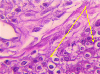
Metritis mononuclear infiltration in myometrium. (400x H & E stain).


