
Ovine Intraorbital Hydatid Cyst


Ovine Intraorbital Hydatid Cyst
Muhammad Kashif Maan1*, Rohma Shabbir2


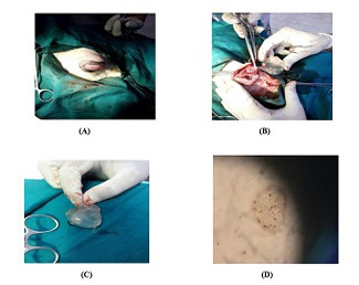
Figure 1
Intraorbital hydatid cyst in 2 year ram (male sheep) with sign of exophthalmos with proptosis. Lid edema was found along with the displacement of the globe on medial side (A). Rhinotomy incision on the outer margins of the eyelid for the removal of the hydatid cyst (B). hydatid cyst after removal from the intra orbital sac by rhinotomy surgery. Cyst is characterized by clear fluid filled pouch with visible scolex inside (C). Microscopic appearance of the hydatid cyst removed from 2-year-old ram. Scolex of the cyst are visible under light microscope examination (D)
March 2022
Vol. 10, Iss. 1, Pages 1-6
