
New and Known Species of Predacious Nematodes of Pakistan
New and Known Species of Predacious Nematodes of Pakistan
Uzma Ishaque1, Nasira Kazi1, Erum Iqbal1* and Shahnaz Dawar2

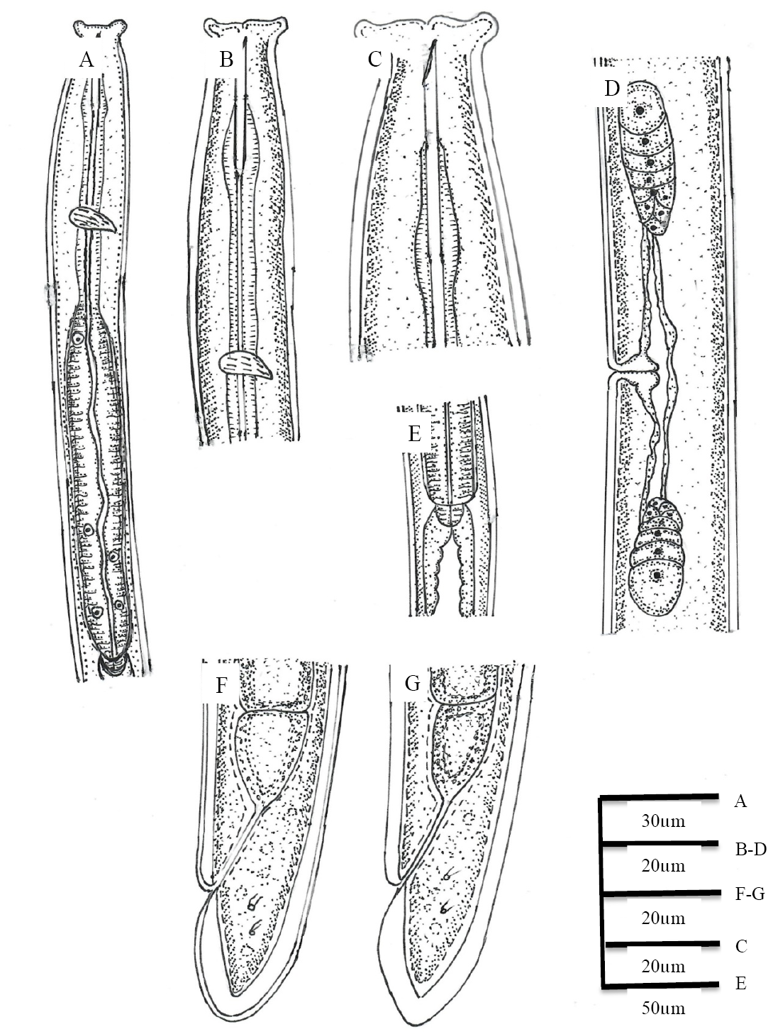
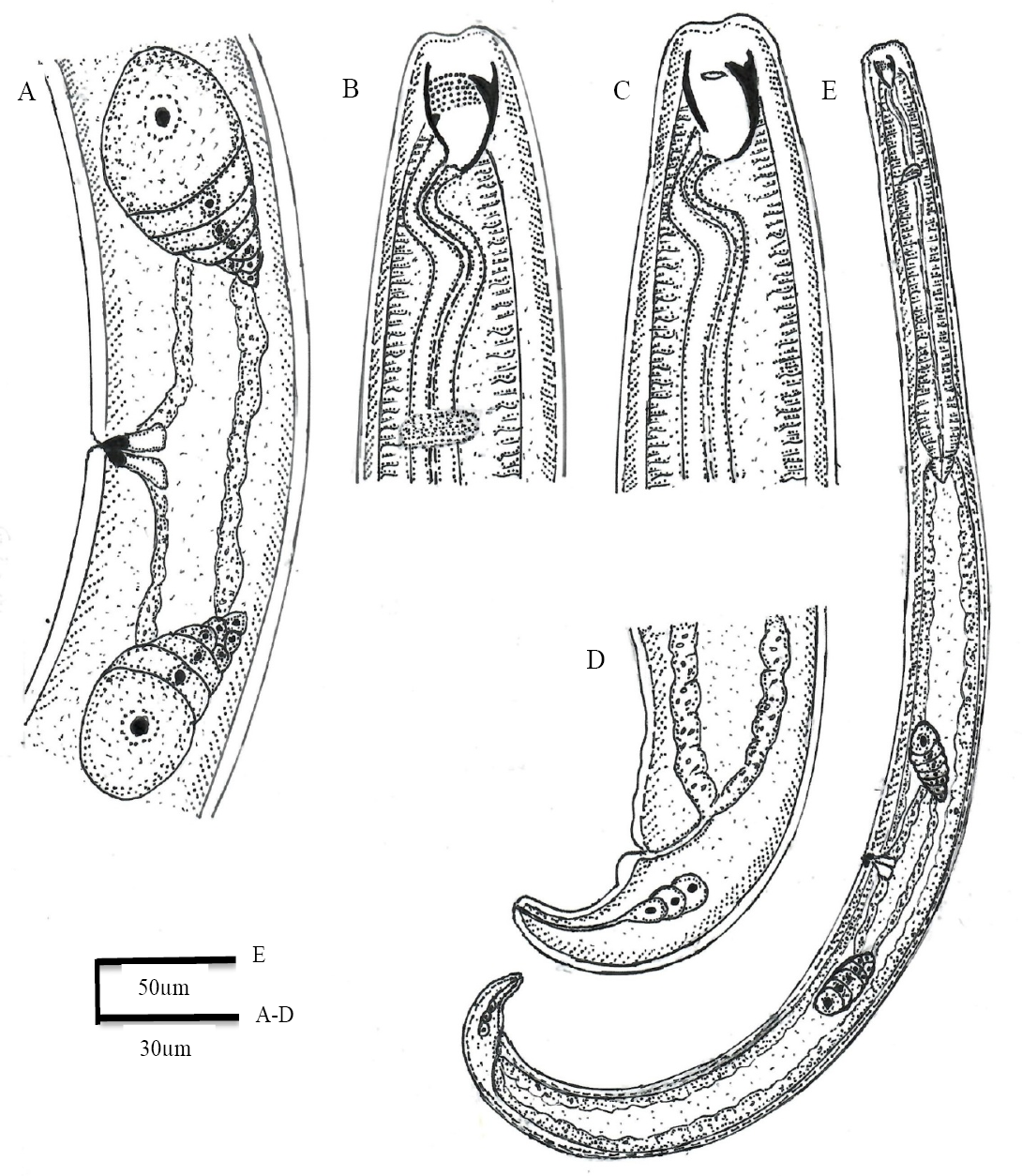
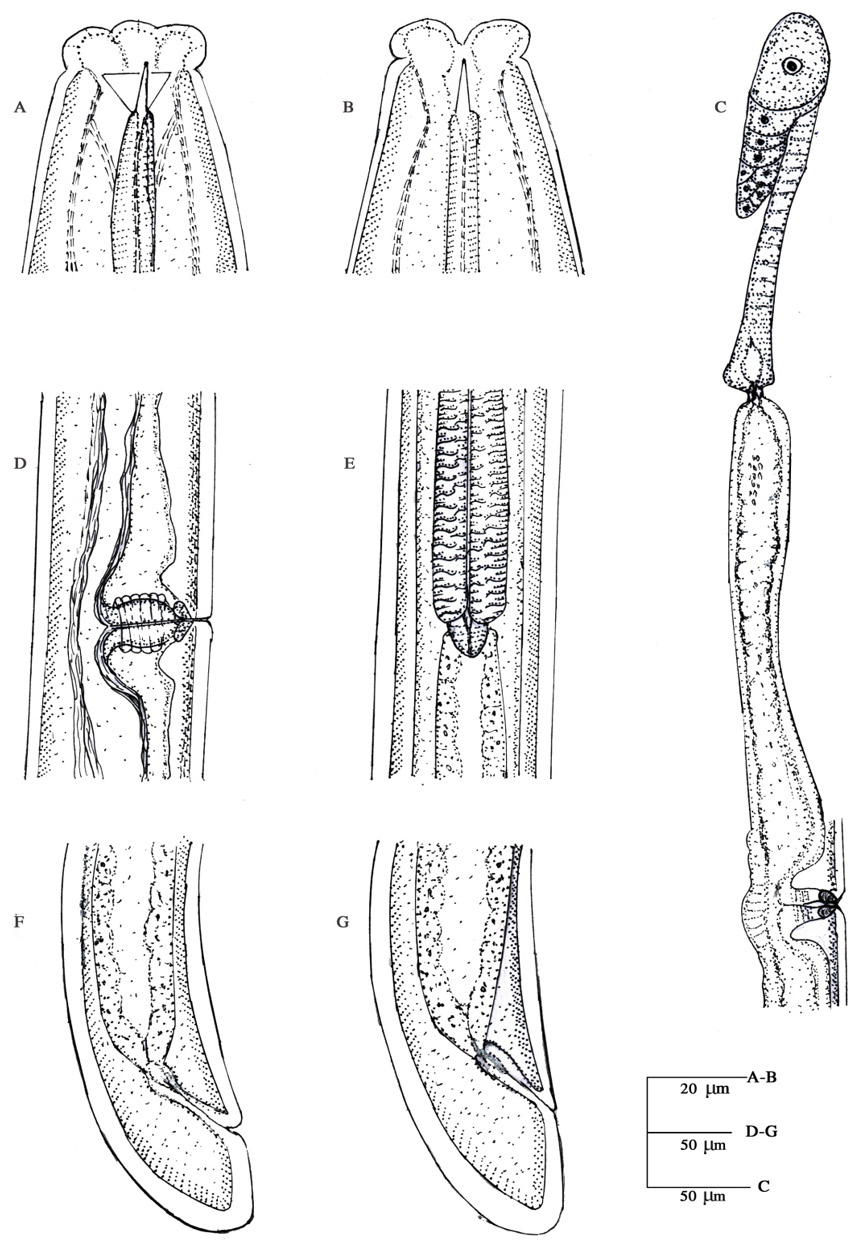
Mylonchulus musae n. sp. Female: A, reproductive system; B, anterior region; C, anterior region showing amphid; D, tail region and E, whole body.
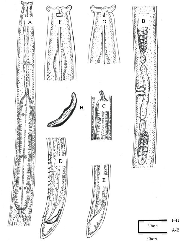
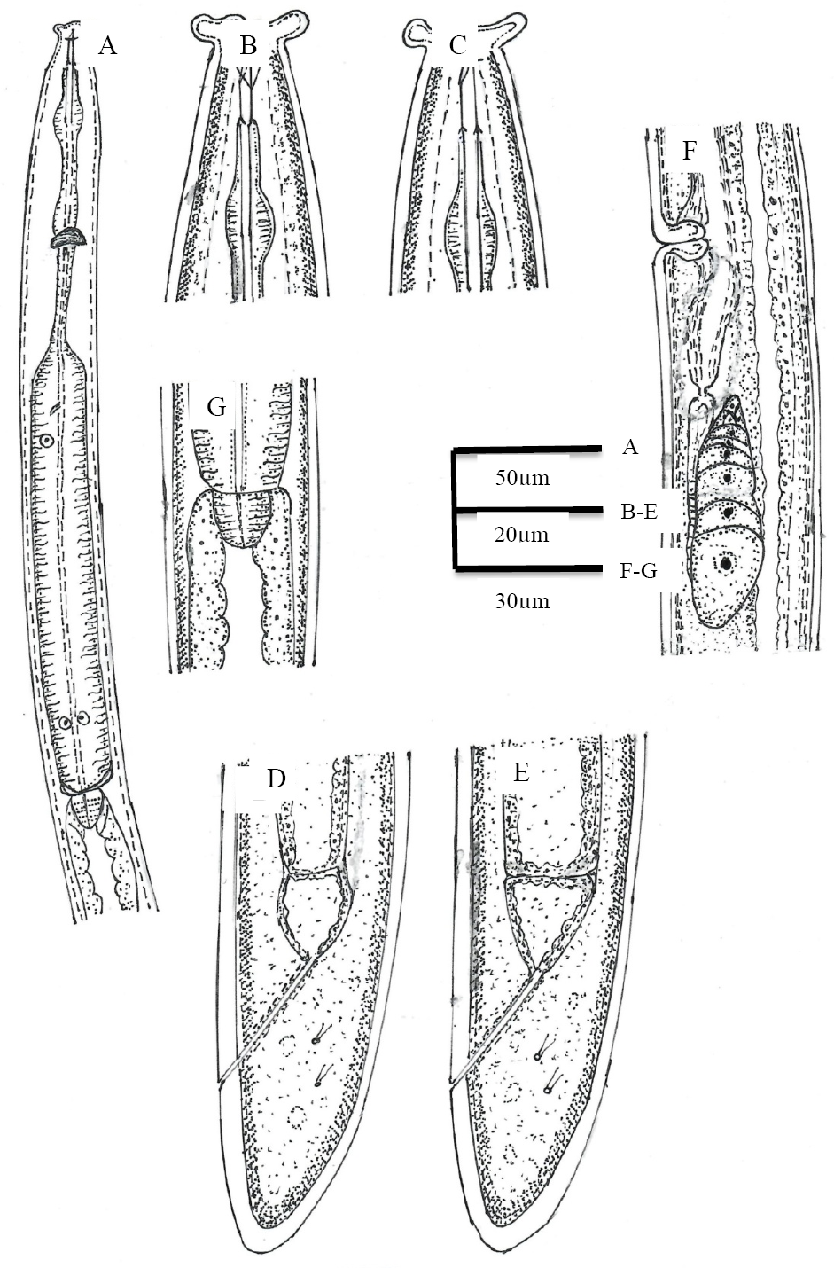
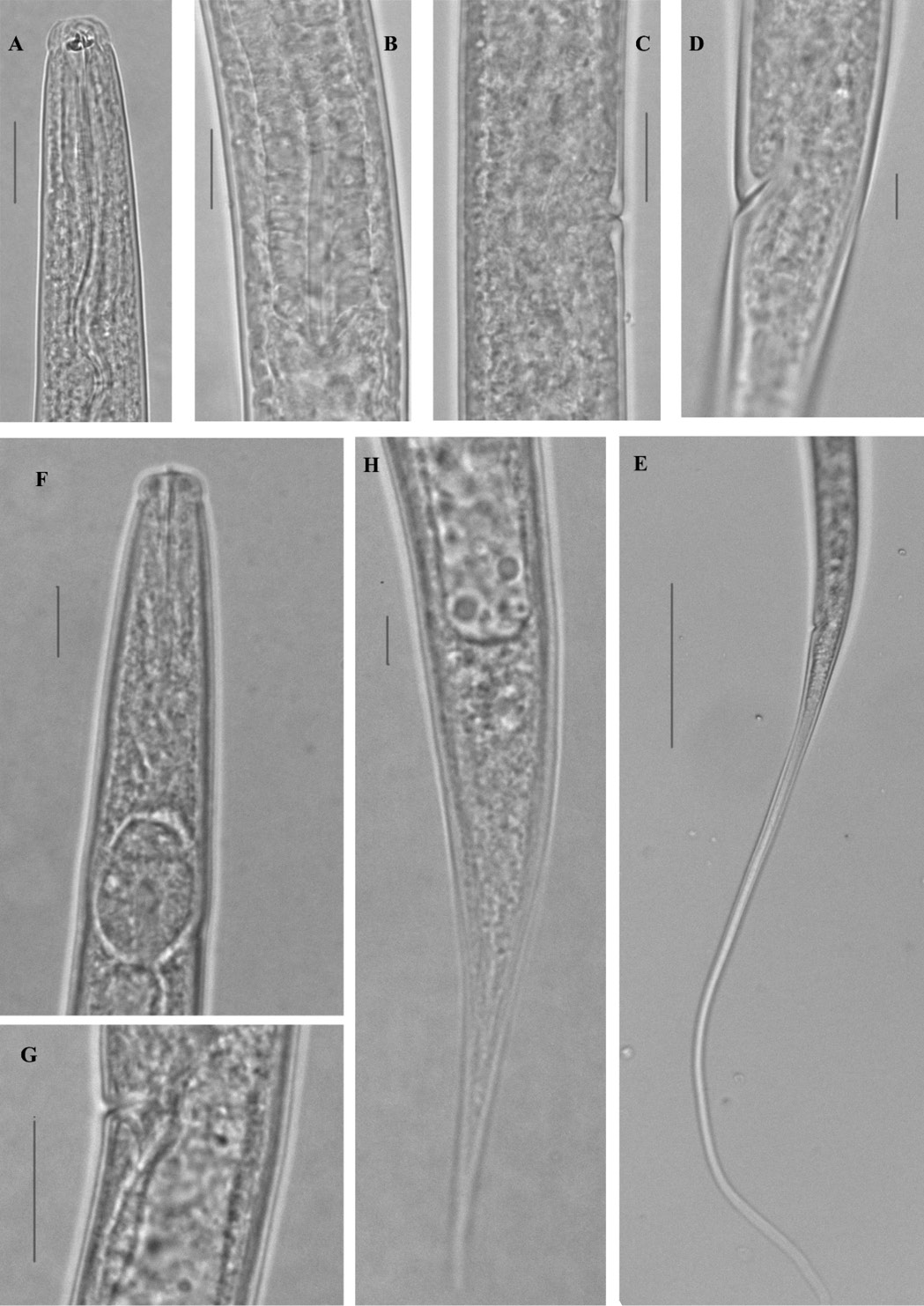
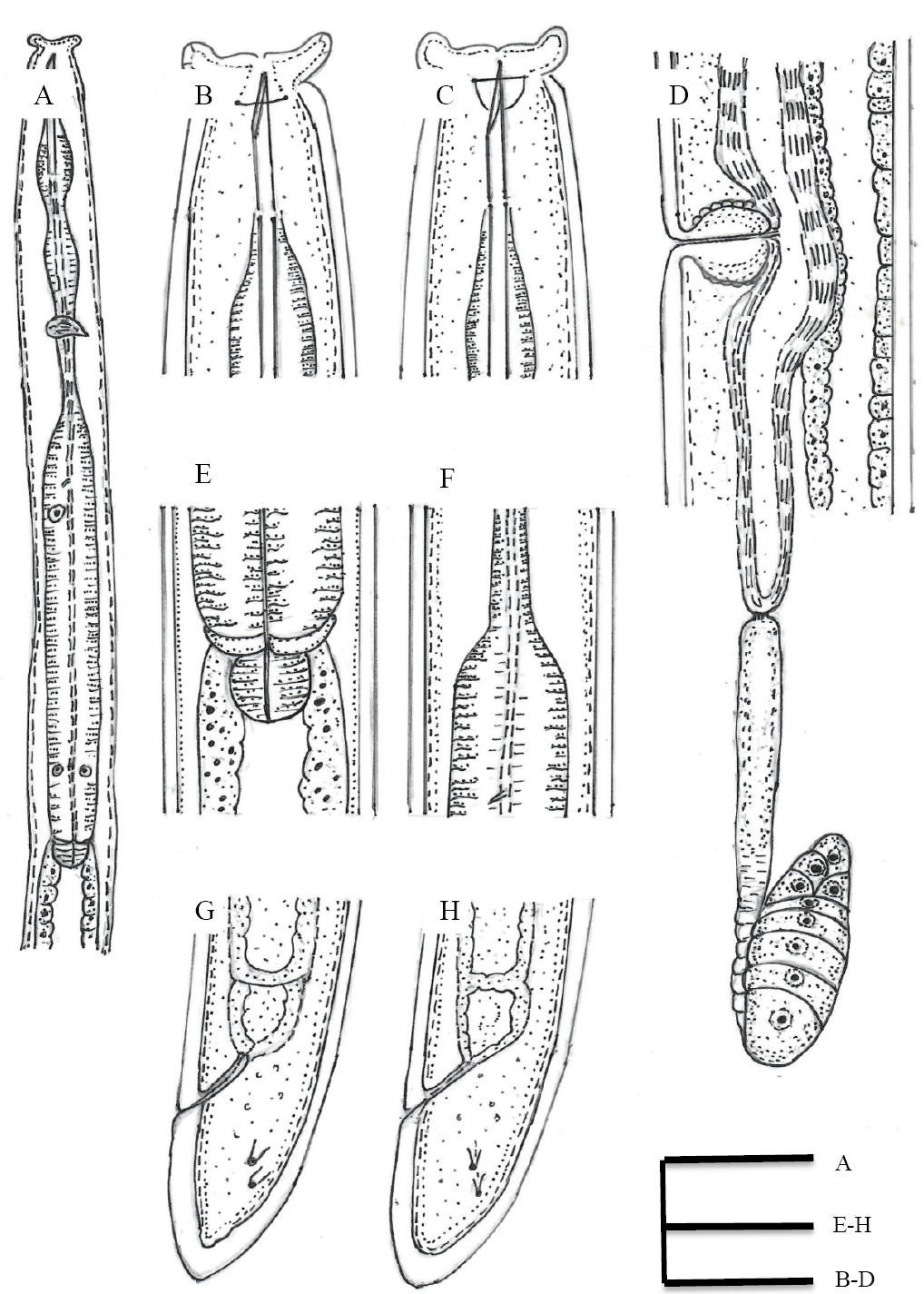
Discolaimus tabacum n. sp. Female: A, Oesophageal region; B, Reproductive system; F, Anterior region; C, Anterior end of oesophageal bulb showing constriction; E, Tail region; Male: G, Anterior region; H, Spicules; D, Tail region showing supplements.
Discolaimus omanensis Siddiqi, 2005. Female: A, Oesophageal region; B, Anterior region showing amphid; C, Anterior region; D, E, Tail region; F, Reproductive system; G, Cardial region.
Discolaimus laksi Khan and Laha, 1982. Female: A, Oesophageal region; B and C, Anterior region; D, Reproductive system; E, Cardial region; F and G, Tail region.
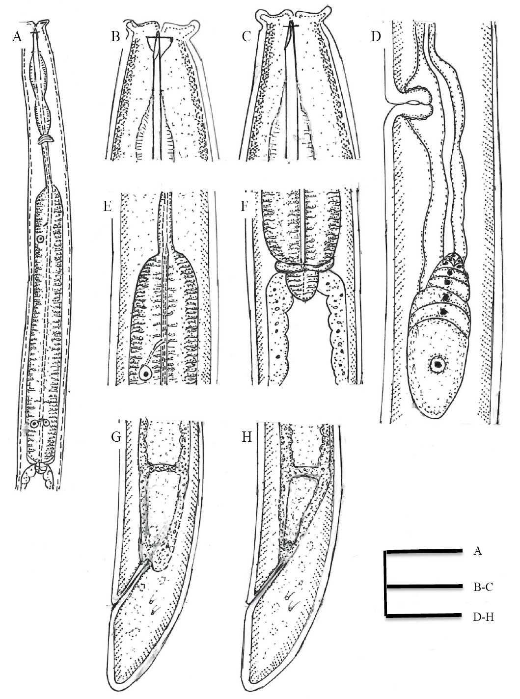
Discolaimus conicus Siddiqi, 2005. Female: A, Oesophageal region; B, Anterior region showing amphid; C, Anterior region; D, Reproductive system; E and F, Anterior and posterior end of oesophageal bulb, respectively; G, H, Tail region.

Discolaimus paratenaxs Siddiqi, 2005. Female: A, Oesophageal region; B, Anterior region; C, Anterior region showing amphid; D, Reproductive system; E and F, Posterior and anterior end of oesophageal bulb, respectively; G and H, Tail region.
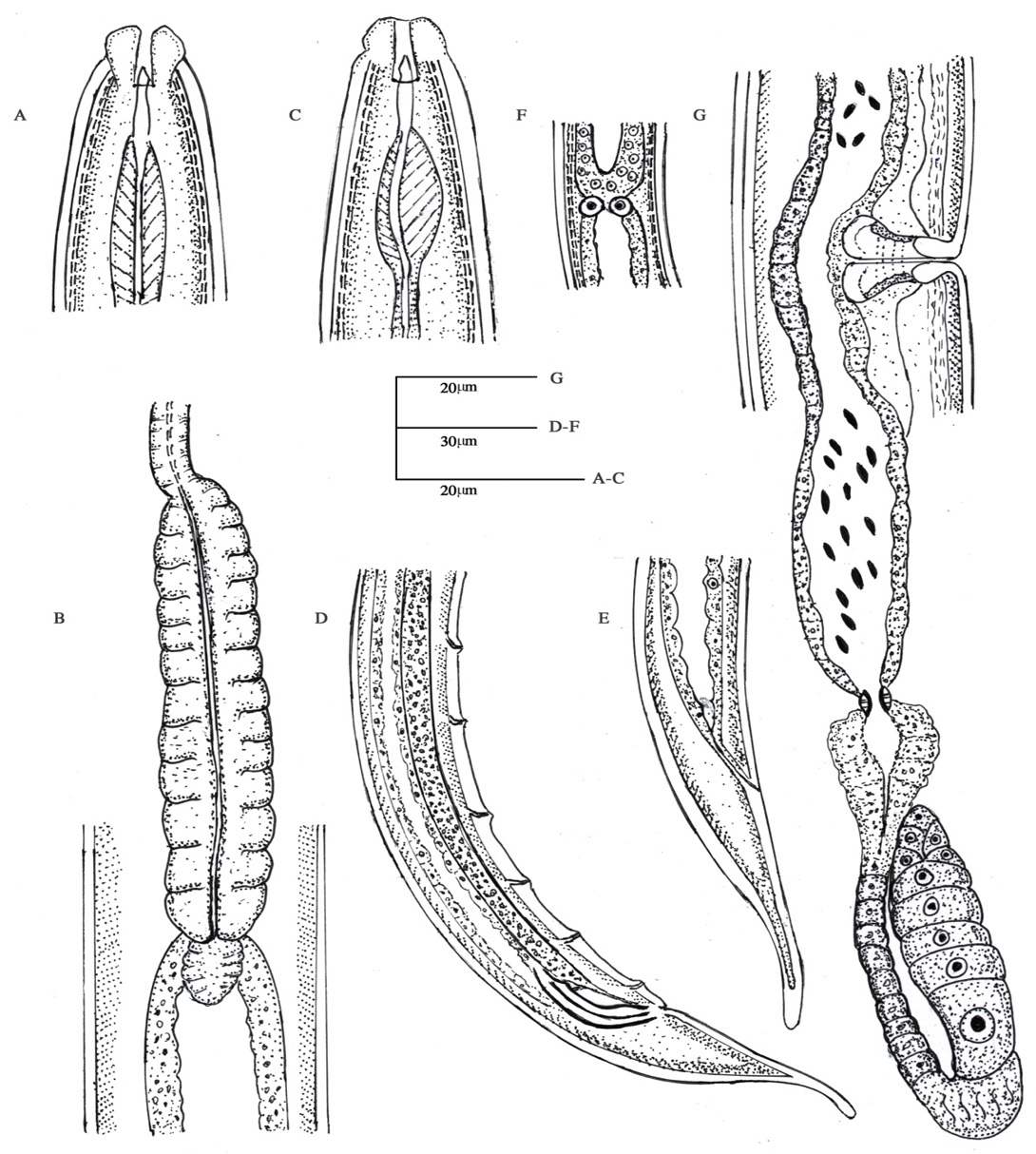
Dorylaimoides micoletzkii (de Man, 1921) Thorne and Swanger, 1936. Female: A, Anterior region; B, Oesophageal bulb; E, Tail region; F, Intestine-prerectum junction; G, Reproductive system; Male: C, Anterior region; D, Tail region showing supplements.

Sectonema ventralis Thorne, 1930. Female: A, Anterior region showing amphid; B, Anterior region; C, Reproductive system; D, Vulval region; E, Cardial region; F and G, Tail region.
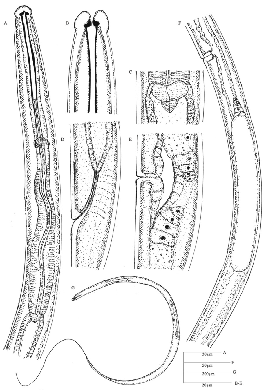
Ironus terranovus Ebsary, 1985. Female: A, Oesophageal region; B, Anterior region; C, Cardial region; D, Rectum; E, Vulval region; F, Reproductive system; G, Whole body.
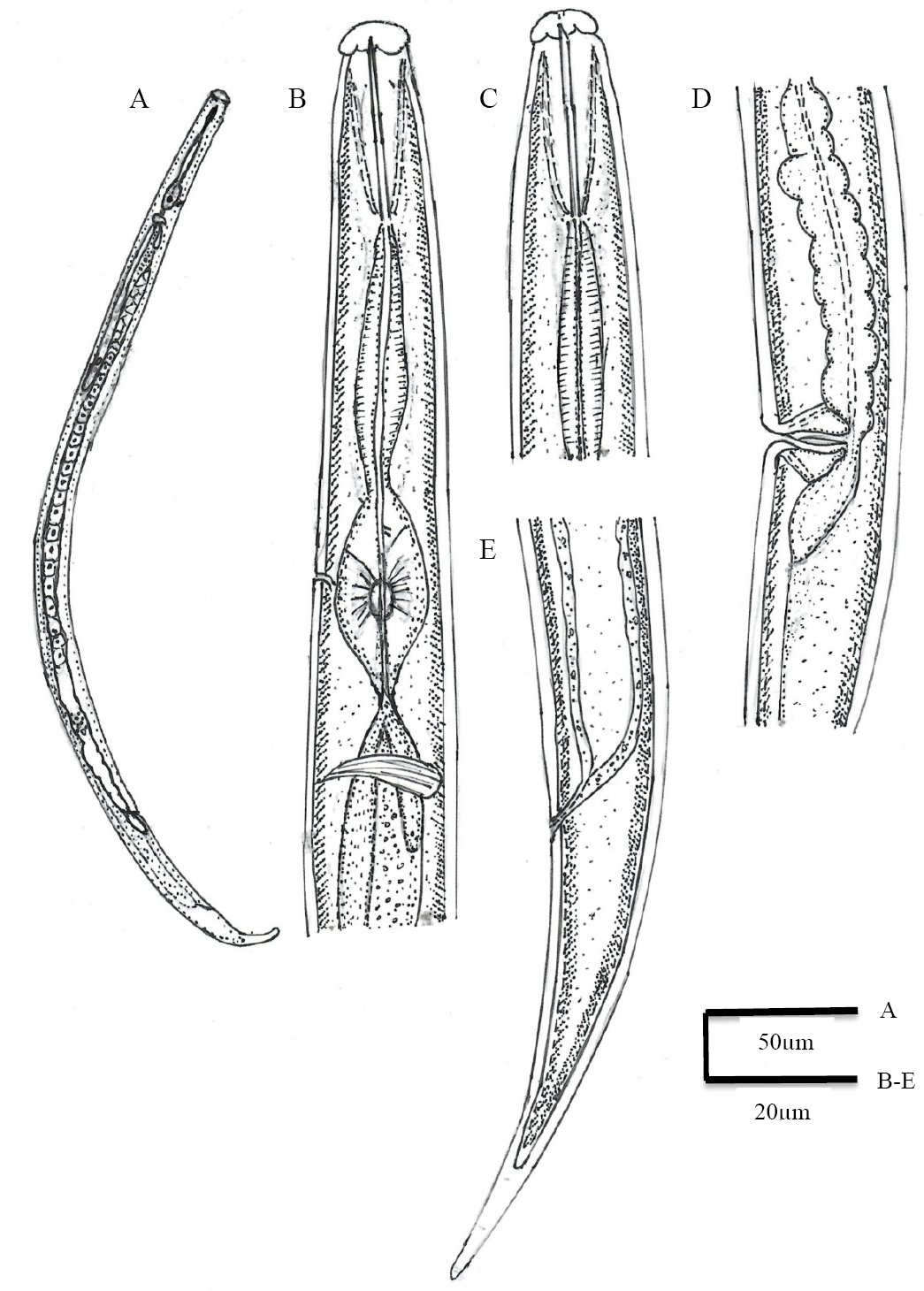
Seinura oswegoensis (Van der Linde, 1938) Goodey, 1960. Female: A, Whole body; B and C, Anterior region; D, Reproductive system; E, Tail region.
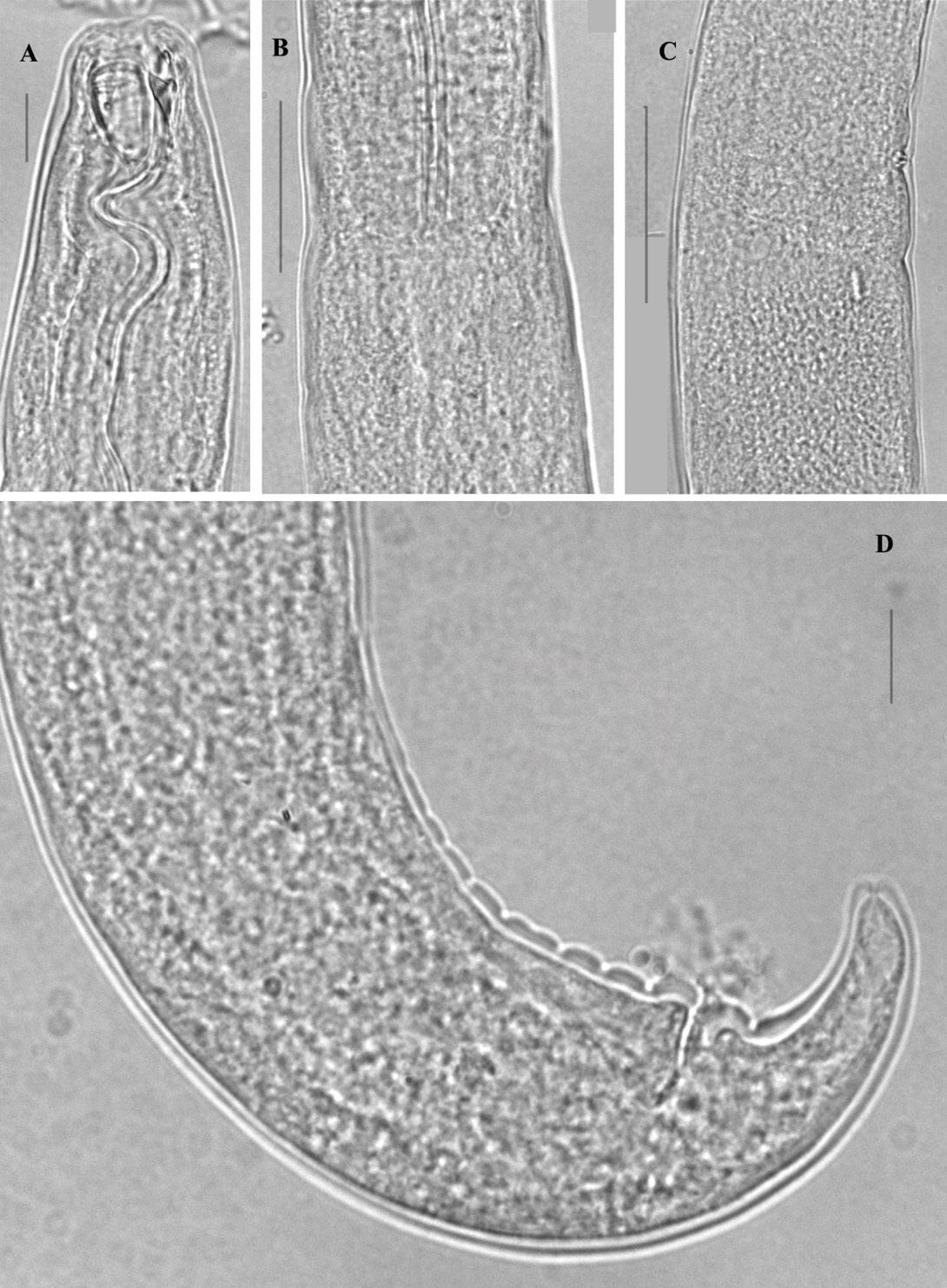
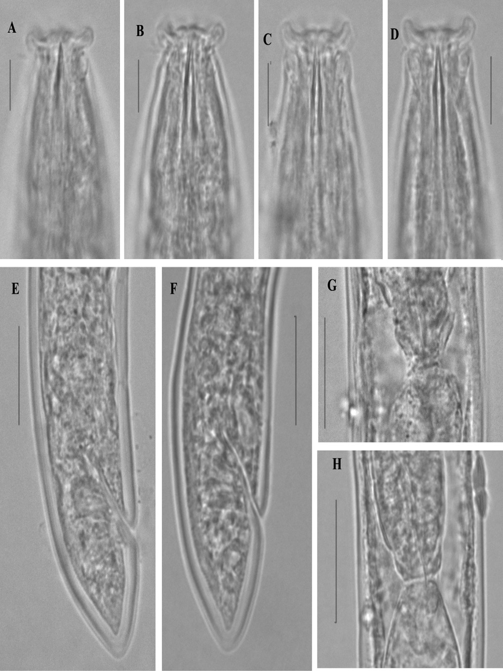
A-D Mylonchulus musae n. sp.

A-H Discolaimus tabacum n. sp.

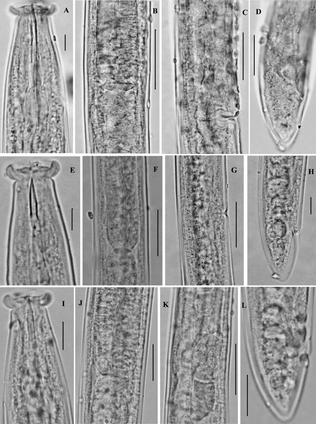
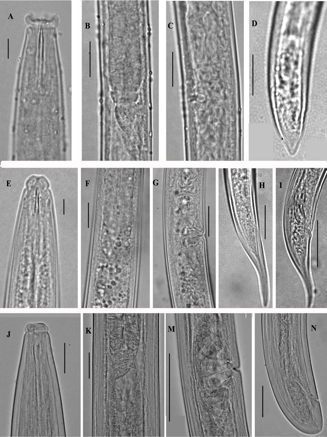
(A-D) Discolaimus conicus Siddiqi, 2005; (E-H) Discolaimus laksi Khan and Laha, 1982; (I-L) Discolaimus omanensis Siddiqi, 2005.

(A-D) Discolaimus paratenaxs Siddiqi, 2005; (E-H) Dorylaimoides micoletzkii (de Man, 1921) Thorne and Swanger, 1936; (I-L) Sectonema ventralis Thorne, 1930.
(A-E) Ironus terranovus Ebsary, 1985; (F-H) Seinura oswegoensis (Van de Linde, 1938) Goodey, 1960

