Morphometric Analysis of Skin in Different Body Regions of Young and Adult Red Sindhi Cattle Bos indicus
Morphometric Analysis of Skin in Different Body Regions of Young and Adult Red Sindhi Cattle Bos indicus
Imran Tarique1*, Muhammad Ghiasuddin Shah1, Illahi Bux Kalhoro1,
Zaheer Ahmed Nizamani2, Mansoor Tariq 2, Jamil Ahmed Gandahi1,
Saqib Ali Fazlani3 and Benazir Sahito3
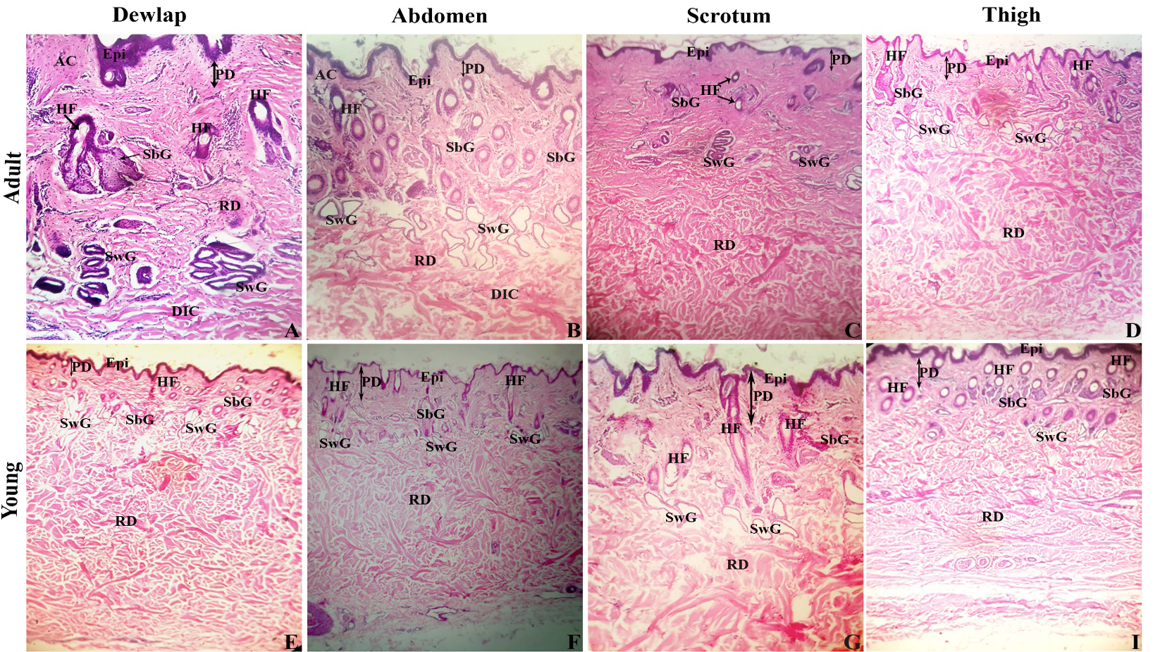
Comparative histological structure of red Sindhi cattle. Epi, epidermis; AC, areolar connective tissue; PD, papillary dermis; RD, reticular dermis; HF, hair follicle; SbG, sebaceous gland; SwG, sweat gland; DIC, dense irregular connective tissue. Scale bar = 100 µm (A-I).
Histological structure of skin appendages of red Sindhi cattle. SbG, sebaceous gland (arrow); SwG, sweat gland (curved arrow); HF, hair follicle (arrowhead); Hc, hair canal; HRS, hair root sheath; Bv, blood vessel.
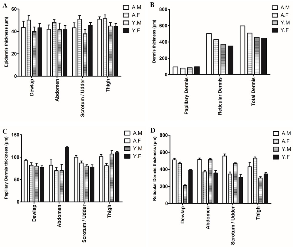
Thickness (µm) of skin layers in different regions of male (adult and young) and female (adult and young) red Sindhi cattle. Data presented as Mean ± SEM. A.M, adult male; A.F, adult female; Y.M, young male; Y.F, young female.
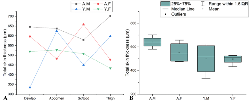
Thickness (µm) of Total skin layer of red Sindhi cattle. A, Comparative curve lines among adult and young genders from dewlap to thigh regions. B, Box plot showing the pattern of quantitative data of adult and young genders.


