Morphological and Morphometrical Comparative Study of Thyroid and Adrenal Glands Between Suckling and Adult Local Male Cats (Felis catus)
Diyar Mohammad Hussein*, Iman Mousa Khaleel
Department of Anatomy, College of Veterinary Medicine, University of Baghdad, Iraq.
*Correspondence | Diyar Mohammad Hussein, Department of Anatomy, College of Veterinary Medicine, University of Baghdad, Iraq; Email: dmh201094@mu.edu.iq
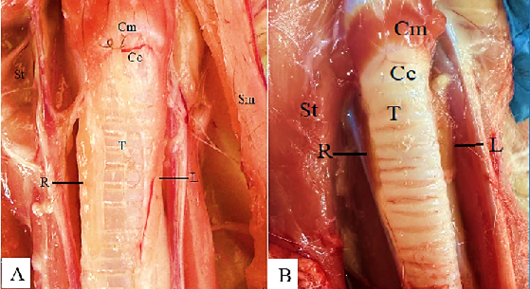
Figure 1:
Photograph of the gross anatomy of thyroid gland in male cats. A: In suckling cats; B: In adult cats.
R, Right lobe; L, Left lobe; T, Trachea; St, Sternothyroideus muscle; Sm, Sternomastoideus muscle; Cc, Cricoid cartilage; JV. Jugular Vein.
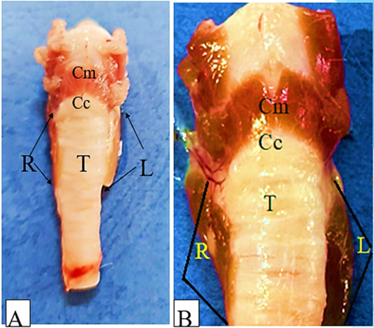
Figure 2:
Photograph of gross anatomy of the thyroid glands in male cats. A, suckling cats; B, adult cats.
R, Right lobe; L, Left lobe; Cc, Cricoid cartilage; T, Trachea; Cm, Cricoid muscle.
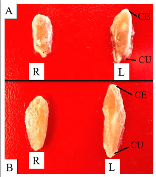
Figure 3:
Photograph of gross anatomy of the thyroid glands in male cats. A, In suckling cats; B, in adult cats.
R, Right lobe; L, Left lobe; CE, cranial end; CU, caudal end.
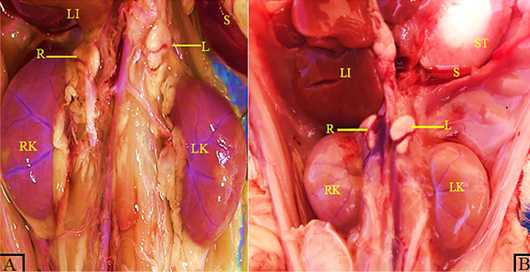
Figure 4:
The gross anatomy of adrenal gland in male cats. A: Suckling cats; B: Adult cats.
R, Right adrenal gland; L, Left adrenal gland; RK, Right kidney; LK, Left kidney; LI, liver; S, spleen; ST, Stomach.
Figure 5:
Photograph of the gross anatomy (external surface) of adrenal glands in male cats. A: Suckling cats; B: Adult cats. R, Right adrenal gland; L, Left adrenal gland.
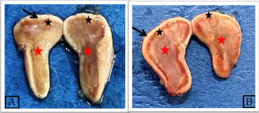
Figure 6:
Photograph of the gross anatomy (internal surface) of right adrenal glands shows. A: In suckling cats: no clear limitation between cortex and medulla, B: n adult cats: clear limitation between cortex (black star) and medulla (Red star), Capsule (black arrow).


