Morphologial and Anatomical Studies of Tea Varieties and Clones Grown at Nthri, Shinkiari, Mansehra, Pakistan
Morphologial and Anatomical Studies of Tea Varieties and Clones Grown at Nthri, Shinkiari, Mansehra, Pakistan
Danish Kamal2, Muhammad Abbass Khan1, Ghulam Mujtaba-Shah1, Naveed Ahmed1, Maryam Iqbal¹, Basharat Hussain Shah1 and Imtiaz Ahmed1*
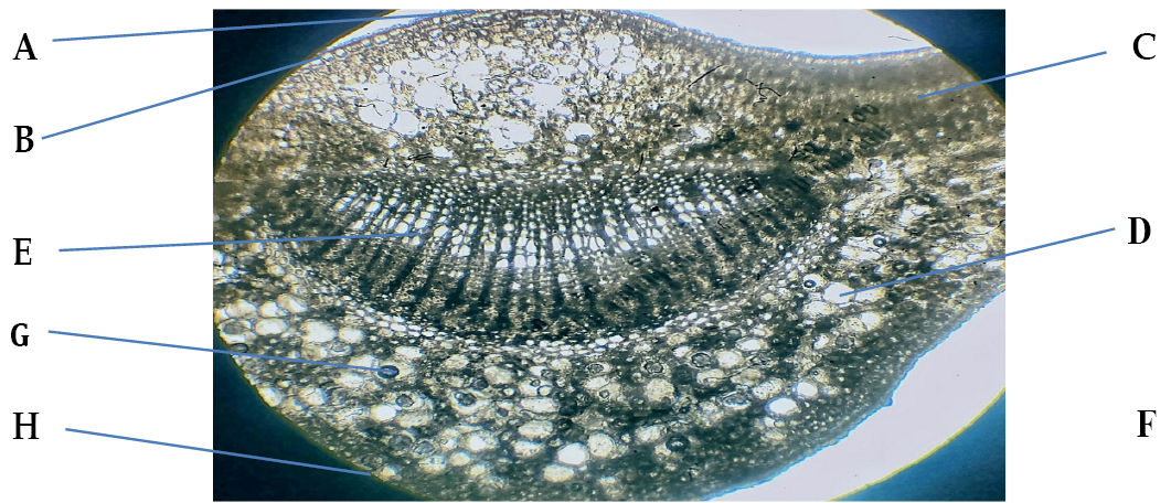
Transverses section of various part of tea leaf: A: cuticle, B: Upper epidermis, C: Palisade parenchyma, D: Spongy parenchyma, E: Vascular bundle, F: Lower epidermis, G: Parenchyma, H: stoma.
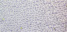
Upper epidermis of tea sample.
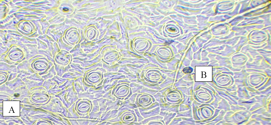
Lower epidermis cell of tea sample: A, Stomata; B, trichome.


