
Molecular Diagnosis of Avihepatovirus a in Naturally Infected Duck Flocks in Egypt


Molecular Diagnosis of Avihepatovirus a in Naturally Infected Duck Flocks in Egypt
A. El-Shemy1*, Hoda M. Mekky2, M. A. Bosila2, A. M. Allam1, Kh. M. Elbayoumi2, M. M. Amer3


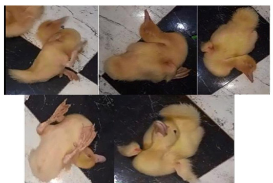
Lateral, keel recumbency, keel over, and retracted head on back.
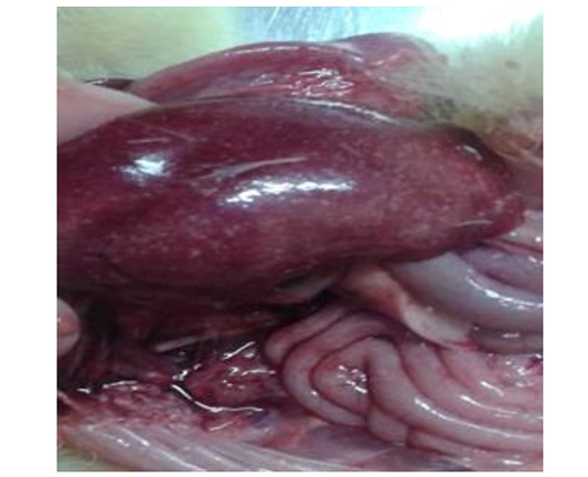
Enlarged liver with hemorrhages.
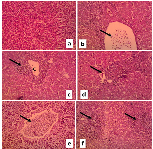
Liver sections of duckling naturally infected with DVH (H&E, X100) showing
a. Normal liver tissue
b. Portal vein and bile duct congestion (arrow) accompanied by hepatic cord disorganization
c. Focal area of coagulative necrosis (C) characterized by inflammatory cells infiltration (arrow) accompanied byhepatic cord disorganization and vacuolar degeneration in the cytoplasm of the hepatocytes
d. Focal area of coagulative necrosis characterized by inflammatory cells infiltration (arrow) accompanied byhepatic cord disorganization and vacuolar degeneration in the cytoplasm of the hepatocytes
e. Diffuse vacuolar degeneration and severe necrosis of hepatocytes and lymphocytic infiltration of the central vein
f. Multifocal area of coagulative necrosis characterized by inflammatory cells infiltration (arrow) accompanied byhepatic cord disorganization and vacuolar degeneration in the cytoplasm of the hepatocytes
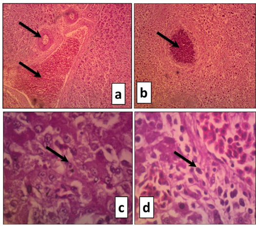
Liver sections of duckling naturally infected with DVH (H&E) showing
a. Severe portal vein congestion (arrow). The bile duct suffered from narrowing because of epithelial lining hypertrophy accompanied by severe hepatocyte necrosis (X100)
b. Focal area of coagulative necrosis characterized by inflammatory cells infiltration (arrow) accompanied byhepatic cord disorganization and severe hepatocytes necrosis (X200)
c. Large eosinophilic intranuclear inclusion surrounded by a clear halo in the hepatocytes (X400)
d. Large eosinophilic intranuclear inclusion surrounded by a clear halo in the hepatocytes (X400)

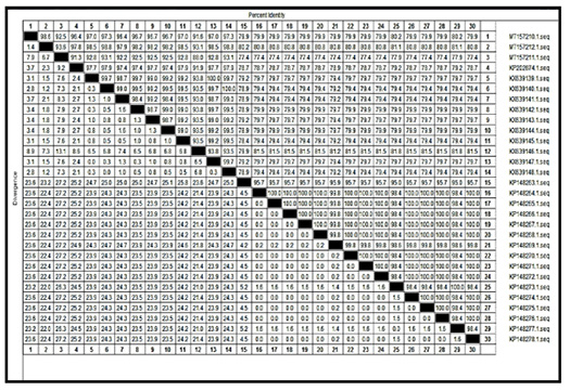
Nucleotide identities of Egyptian isolates with selected published field Egyptian strain references and vaccinal DHAV-1 sequences of different serotype.

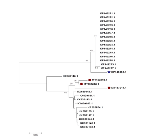
Phylogenetic tree based on a partial sequence of the DHAV-1 3D gene. The dots refer to viruses isolated in the current study, and the triangle refers to the vaccine strain. The robustness of individual nodes of the tree was assessed using 1,000 replications of bootstrap resampling of the originally aligned nucleotide sequences
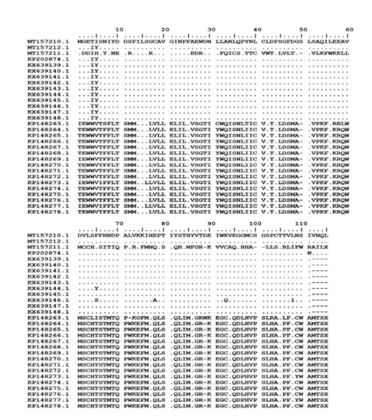
Deduced amino acid sequence of the 3D gene of the isolated strainscompared with the 3D gene of thepublished vaccinal and field Egyptian strains

