Molecular Detection of Methicillin-resistant Staphylococcus aureus (MRSA) from a Clinical Case of Myiasis Wound
Molecular Detection of Methicillin-resistant Staphylococcus aureus (MRSA) from a Clinical Case of Myiasis Wound
Pravin Mishra1, Md. Muket Mahmud2, Md. Ahosanul Haque Shahid2, Alamgir Hasan2, Vivek Kumar Yadav3 and Moinul Hasan1*
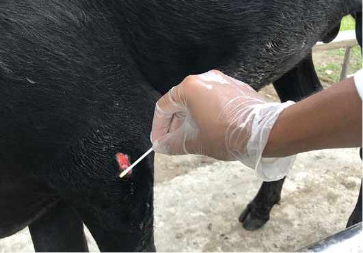
Collection of swab sample from myiasis wound.
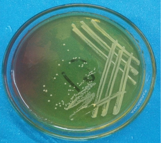
Fermentation of MSA by Staphylococcus aureus indicated by Smooth and convex formation of a yellowish colony.
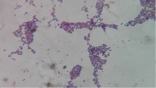
Staining characteristics of bacteria: Grape like clusters were found which are the characteristics of Staphylococcus aureus.
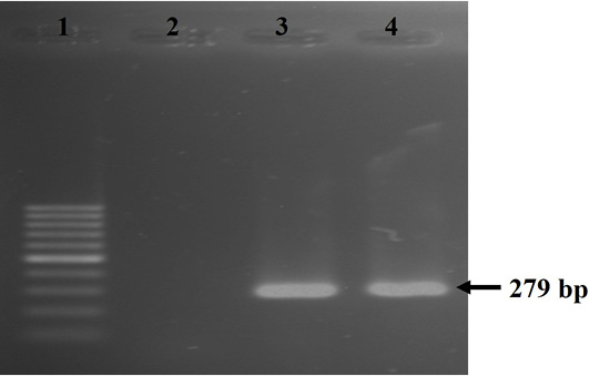
Results of PCR of Staphylococcus aureus specific nuc gene (size=279 bp). Here, Lane 1: 100 bp ladder, Lane 2: negative control, Lane 3: positive control, Lane 4- amplified nuc gene of S. aureus.
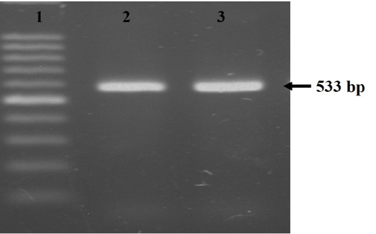
Amplification of the mecA gene (size=533 bp). Here, Lane 1: 100 bp ladder, Lane 2: positive control, Lane 3: amplified mecA gene of Staphylococcus aureus.


