MRI Investigations on Venomous Glands of Brown Bullhead, Ameiurus nebulosus (Lesueur, 1819) (Actinopterygii: Ictaluridae)
MRI Investigations on Venomous Glands of Brown Bullhead, Ameiurus nebulosus (Lesueur, 1819) (Actinopterygii: Ictaluridae)
Daniel Cocan1, Vioara Mireşan1, Florentina Popescu1, Radu Constantinescu1, Aurelia Coroian1, Călin Laţiu1, Romulus Valeriu Flaviu Turcu3, Alexandru Ştefan Fărcăşanu3 and Cristian Martonos2,*
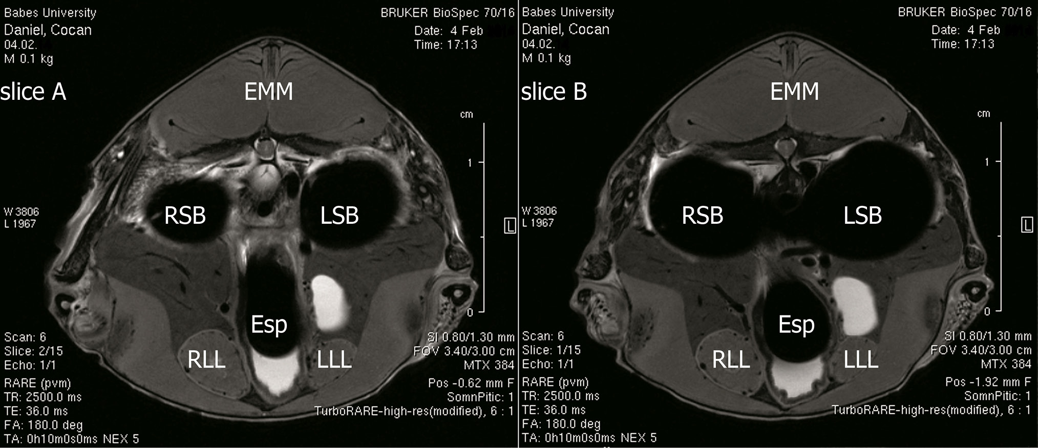
Axial section of brown bullhead (Ameiurus nebulosus). Slice scanned cranial to the first dorsal fin. A, slice 2/15; B, slice 1/15.
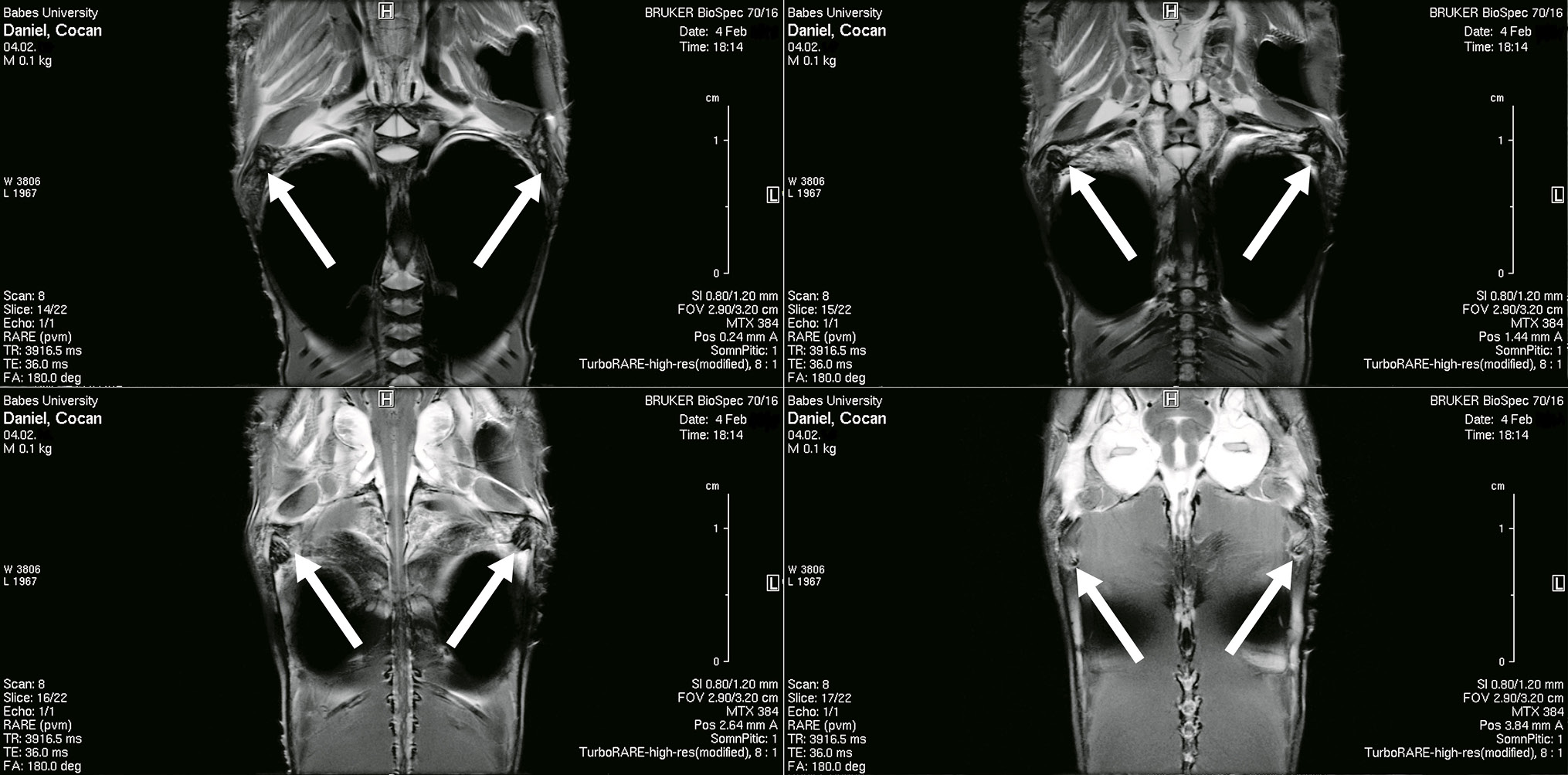
Venomous pectoral glands of Brown bullhead (Ameiurus nebulosus). Different aspects in successive slices from ventral to dorsal part (section in coronal plane).
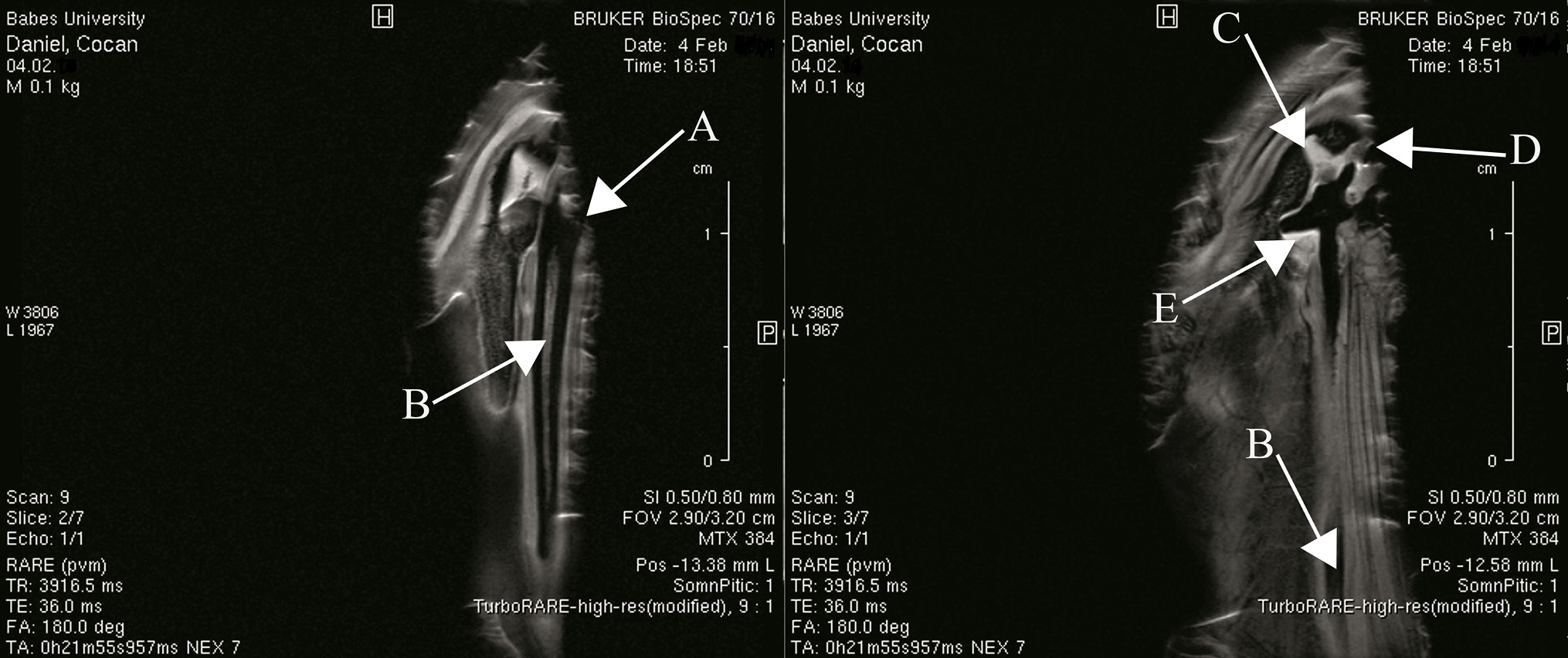
Morphology of venomous pectoral glands of Brown bullhead (Ameiurus nebulosus) in sagittal section. A, venomous pectoral gland; B, pectoral spine; C, anterior process; D, dorsal process; E, ventral process.
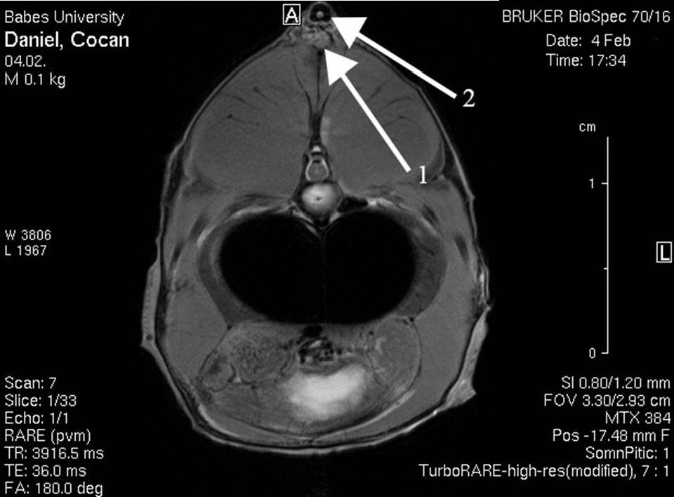
Anatomical topography of venomous dorsal gland in axial section. Arrow 1, venomous dorsal gland; Arrow 2, dorsal spine.


