
Lithium Induced Histological and Thyroid Hormone Alterations Observed in Channa punctatus and Oreochromis niloticus


Lithium Induced Histological and Thyroid Hormone Alterations Observed in Channa punctatus and Oreochromis niloticus
Selvaraj Thanga Malathi¹, Venkatraman Anuradha1*, Mohamed Yacoob Syed Ali2, Muhameed Sajjad Sarwar3, Nagarajan Yogananth2

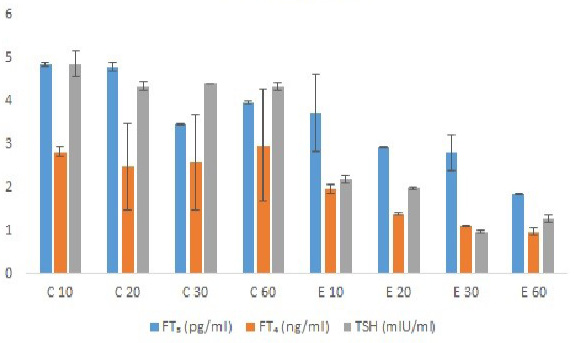
Changes in free thyroid hormones levels in blood of Channa punctatus exposed to sublethal concentration (30%) of Lithium in different exposure days. Values are expressed as mean ± SD for above observation. – or + indicate percent decrease or increase over control. For two-way ANOVA P-values mentioned as: P>0.05 NS; FT4- P> 0.05 NS; TSH- P> 0.05 NS.

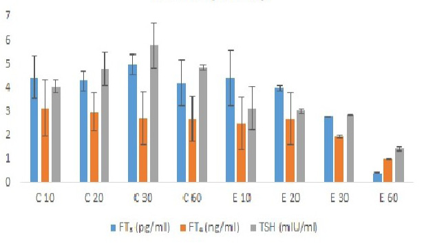
Changes in free thyroid hormones levels in blood of Oreochromis niloticus exposed to sublethal concentration (30%) of Lithium in different exposure days. Values are expressed as mean ± SD for above observation. – or + indicate percent decrease or increase over control. For two-way ANOVA P-values mentioned as: P>0.05 NS; FT4- P> 0.05 NS; TSH- P> 0.05 NS.
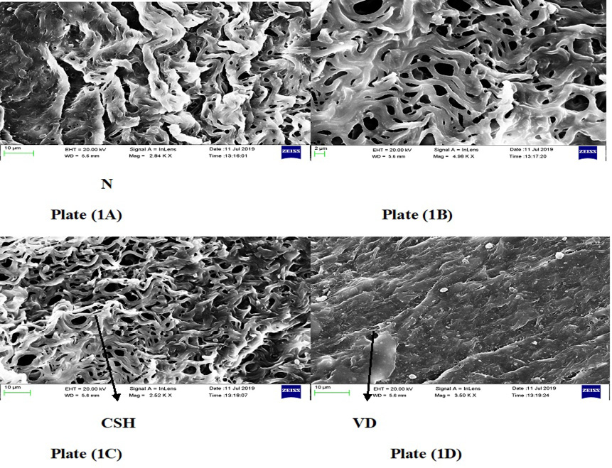
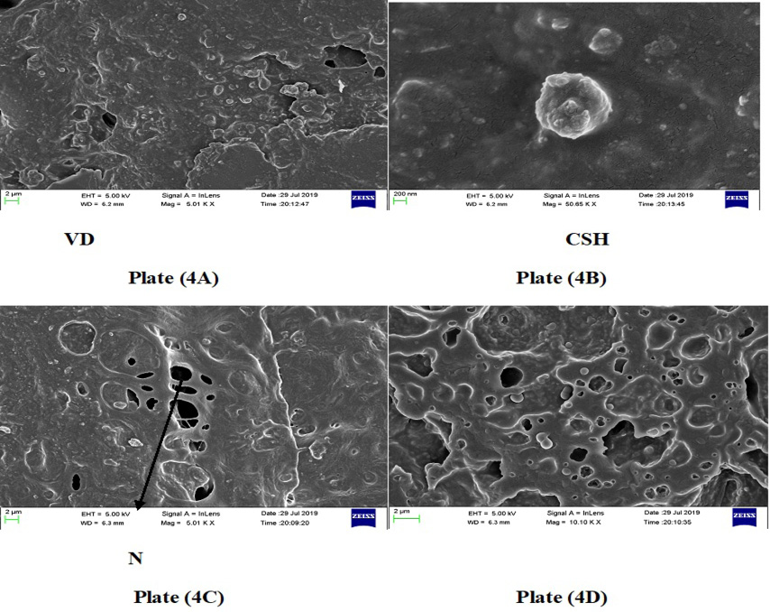
FESEM (field emission scanning electron microscopy) of liver tissues in the group of Channa punctatus treated with lithium chloride. CSH, cloudy swelling of hepatocytes; VD, vascular degeneration and N, necrosis. (Upon 30 days exposure of lithium).
FESEM images of liver tissue in the group of Channa punctatus treated with lithium chloride. CSH, cloudy swelling of hepatocytes; VD, vascular degeneration and N, necrosis. (Upon 60 days exposure of lithium).
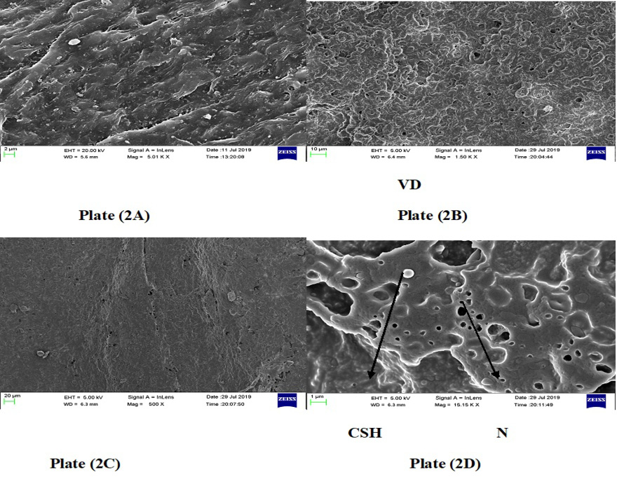
FESEM imaged of liver tissue in the group of Oreochromis niloticus treated with lithium chloride. CSH, cloudy swelling of hepatocytes; VD, vascular degeneration and N, necrosis. (Upon 30 days exposure of lithium).

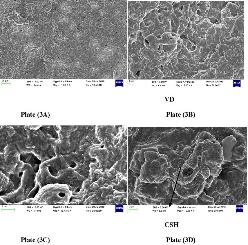
FESEM of liver tissue in the group of Oreochromis niloticus treated with lithium chloride. CSH, cloudy swelling of hepatocytes; VD, vascular degeneration and N, necrosis. (Upon 60 days exposure of lithium).
