
Impact of Drinking Saline Water on Meat Production and Muscles Structures of Barki Lambs


Impact of Drinking Saline Water on Meat Production and Muscles Structures of Barki Lambs
Mohamed Ali Zayed

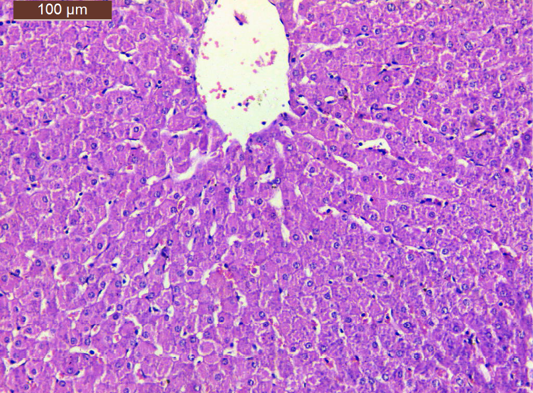
A micrograph of a section liver from lamb drinking fresh water showing the hepatic lobule. Notice the central vein (arrow), sinusoids (red arrow) and hepatocytes cords (yellow arrow) that associated with central nuclei (green arrow) (H and E stain, Scale Bar: 100 µm).

A micrograph of a section liver from lamb drinking fresh water showing collagen fibers around portal vein (red arrow), bile ductile (black arrow), hepatic artery (blue arrow) and lymphatics (arrowhead) (H and E stain, Scale Bar: 100 µm).
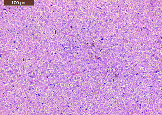
A micrograph of a section liver from lamb drinking saline water showing disturbance of the hepatic lobule. Notice hedropic degeneration in the hepatocytes (H and E stain, Scale Bar: 100 µm).
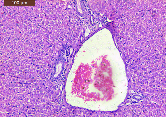
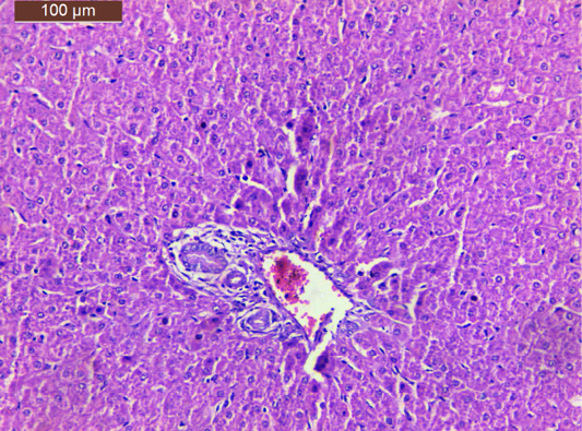
A micrograph of a section liver from lamb drinking saline water showing congested portal tract that associated with inflammatory infiltration (H and E stain, Scale Bar: 100 µm).
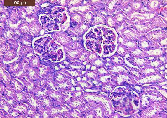
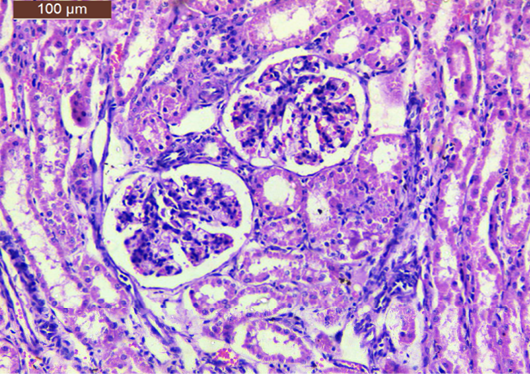
A micrograph of a section kidney from lamb drinking fresh water showing normal renal corpuscles and tubules (H and E stain, Scale Bar: 100 µm).
A micrograph of a section kidney from lamb drinking saline water showing partially degeneration of the glomeruli or atrophy (H and E stain, Scale Bar: 100 µm).

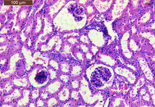
A micrograph of a section kidney from lamb drinking saline water showing lobuolated glomeruli associated with inflammatory infiltration. Note the degeneration of reanal tubules and an interstitial inflammation (H and E stain, Scale Bar: 100 µm).

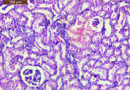
A micrograph of a section kidney from lamb drinking saline water showing interstitial hemorrhage. Notice the cell debris in the lumen of the renal tubules (H and E stain, Scale Bar: 100 µm).

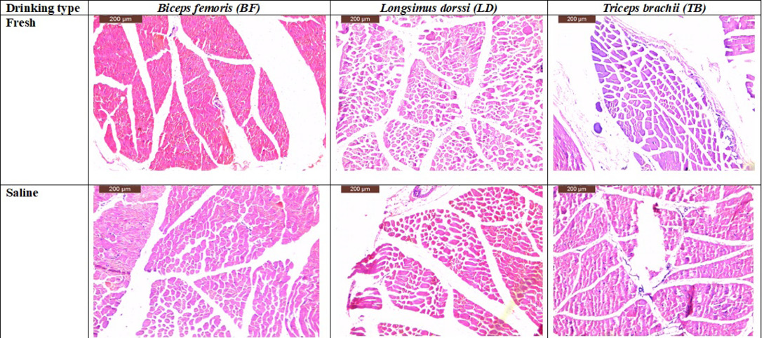
A micrograph of a section Biceps Femoris (BF) from lamb drinking fresh water showing intact muscle fibers however Longsimus Dorssi (LD) and Triceps Brachii (TB) muscles from lamb drinking fresh water showing normal muscle fibers shape in the other hand A micrograph of a section Biceps Femoris muscle from lamb drinking saline water showing relative atrophy of muscle fibers while Longsimus Dorssi muscle from lamb drinking saline water showing relative hypertrophy of muscle fibers moreover Triceps Brachii muscle from lamb drinking saline water showing an elongation of muscle fibers and narrow of the thickness as compared with the normal one (H and E stain, Scale Bar: 200 µm).


