

Histological and Electron Microscopical Structure of Tongue and Lingual Papillae of Guinea Fowl (Numida meleagris)


Histological and Electron Microscopical Structure of Tongue and Lingual Papillae of Guinea Fowl (Numida meleagris)
Ramazan İlgün1*, Nilgün Kuru2, Ferhan Bölükbaş3 and Fatih Mehmet Gür4

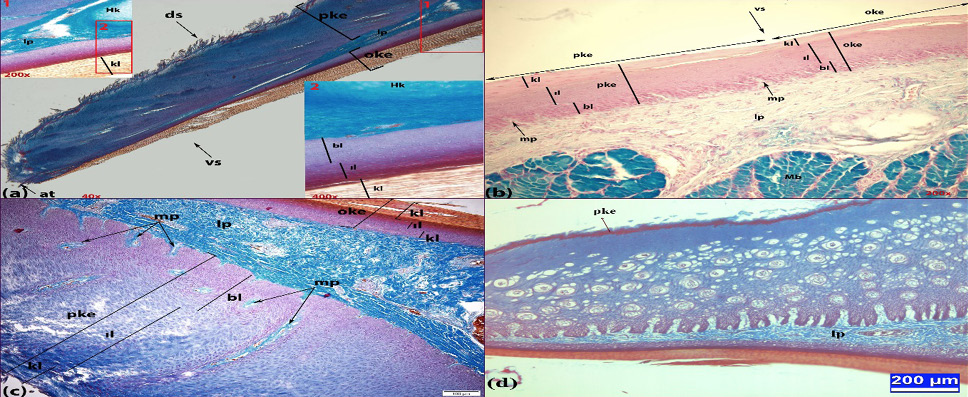
(a) Apex-corpus view of the tongue ( 6-7 weeks). Triple stain 40x; 200x; 400x. (b) Corpus- radix view ventral surface of the tongue (studs). Alcian blue stain 200x. (c) Apex-corpus view of the tongue (studs). Triple stain 100x. (d) Apex view of the tongue (9-13 weeks). Triple stain, Scale bars: 200µm. vs)ventral surface of the tongue; ds) dorsal surface of the tongue; at) apex of tongue; oke) orthokeratinized stratified epithelium; pke) parakeratinized epithelium; lp) lamina propria; bl)basal layer; ıl)intermediate layer; kl)keratinized layer; Hk) Hyaline cartilage; mp) microscobic papillae; Mb)Mucous glands.
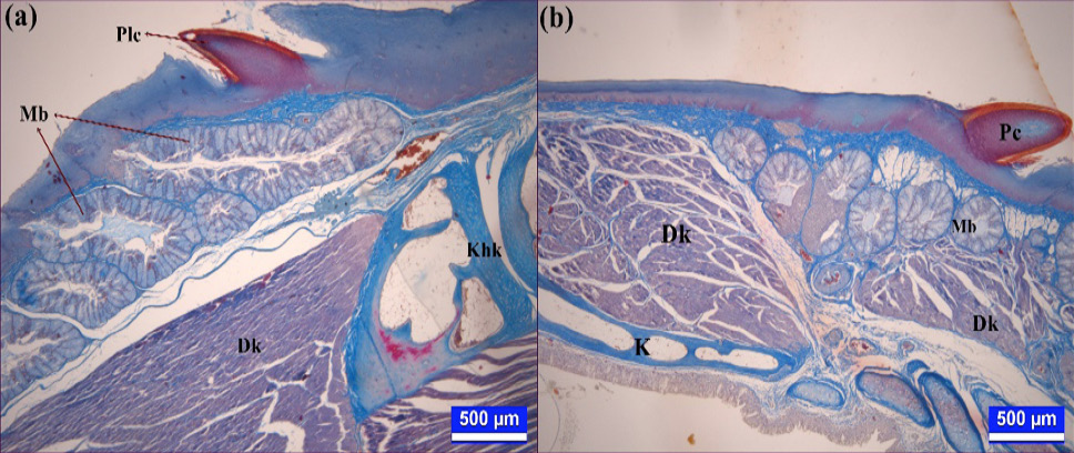
(a) Corpus-radix view of the tongue (9-13 weeks). Triple stain. Scale bars: 500µm. (b) Radix view of the tongue (9-13 weeks). Triple stain. Scale bars: 500µm. Plc) papilla linguales caudales, Mb) mucous glands, Dk) tongue muscle, Khk) hyaline cartilage, Pc) papilla conicae, K) hyaline cartilage.
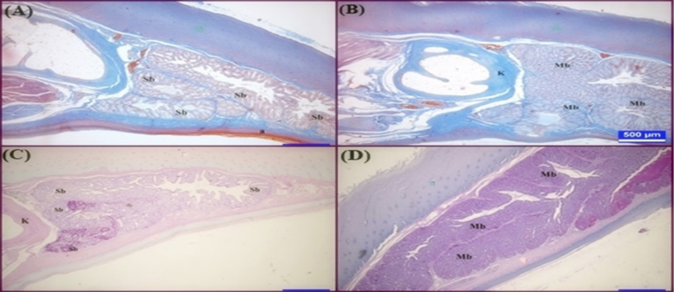
(A) Apex-corpus view of the tongue ( 6-7 weeks). Triple stain. Scale bars: 500µm. , (B) Apex-corpus view of the tongue in the stud, Triple stain. Scale bars: 500µm. (C) Apex view of the tongue ( 6-7 weeks). PAS. Scale bars: 500µm. (D) Mucous glands of the stud. PAS. Scale bars: 500µm. Sb) Serous glands, a) parakeratinized epithelium, Mb) Mucous glands, K) Hyaline cartilage.

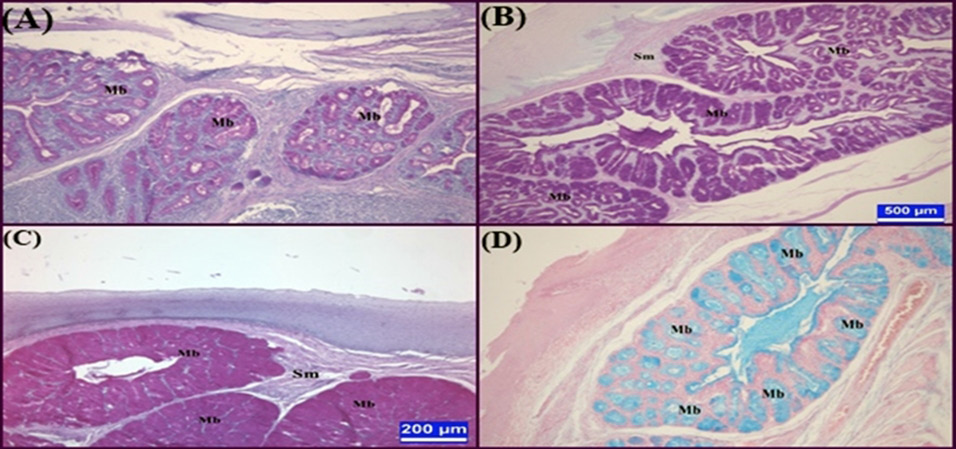
(A) Corpus- radix view of the tongue (6-7 weeks) PAS. Scale bars: 200µm.
The glandular epithelial cells in the corpus and radix regions show a PAS positive reaction at varying degrees of age, Mb) Mucous glands. (B) Corpus-radix view of the tongue (9-13 weeks). PAS. Scale bars: 500µm. The glandular epithelial cells in the corpus and radix regions show a PAS positive reaction at varying degrees of age. Mb). Mucous glands (C) Corpus-radix view of the tongue (stud). PAS. Scale bars: 500µm. The glandular epithelial cells in the corpus and radix regions show a PAS positive reaction at varying degrees of age. Mb) Mucous glands (D) Corpus- radix view of the tongue (6-7 weeks). Alcian blue stain. Scale bars: 200µm. Mb) Mucous glands.

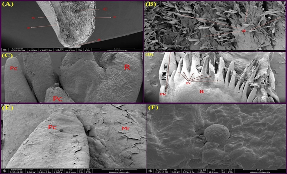
(A) SEM view of the dorsal cross section of the apex part of the tongue in the stud. (a) Stratified Squamous Epithelium (Keratinized), (K) Keratin, (A) Apex, (C) Corpus. (B) A higher magnification of the tongue apex with the stratified squamous epithelium cells, (a) Stratified Squamous Epithelium (Keratinized), (C) SEM view of the papillae of the radix part of the tongue (9-13 weeks). (R) Radix, (Plc) Papilla linguales caudales, (Pc) Papilla conicae. (D) SEM view of the papillae of the radix part of the tongue (stud). (R) Radix, (Plc) Papilla linguales caudales, (Pc) Papilla conicae. (E) SEM view of the papillae of the corpus-radix part of the tongue (9-13 weeks). (Pc) Papilla conicae, (Mr) Epithelial folds, (F) SEM view of the surface of the tongue body and papilla linguales (stud).

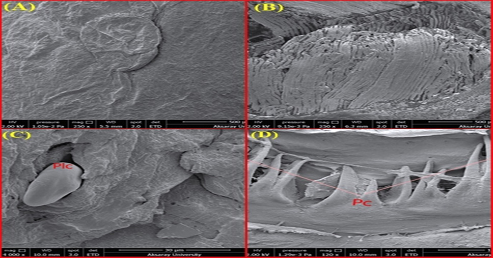
(A) SEM view of the surface epithelium of the corpus portion of the tongue (9-13 weeks). (B) Cross-sectional SEM view of longitudinal tongue muscles (6-7 weeks), (C) SEM view of the surface of the tongue body and papilla linguales caudales (6-7 weeks), (D) SEM view of the papillae of the radix part of the tongue (6-7 weeks), (Pc) Papilla conicae.


