

Histological Structure and Mast Cell Distribution of Hedgehog's Small Intestine


Histological Structure and Mast Cell Distribution of Hedgehog's Small Intestine
Youbao Zhong1, Xianlai Zhang1, Xiaofen Hu1 and Yong Li1, 2,*

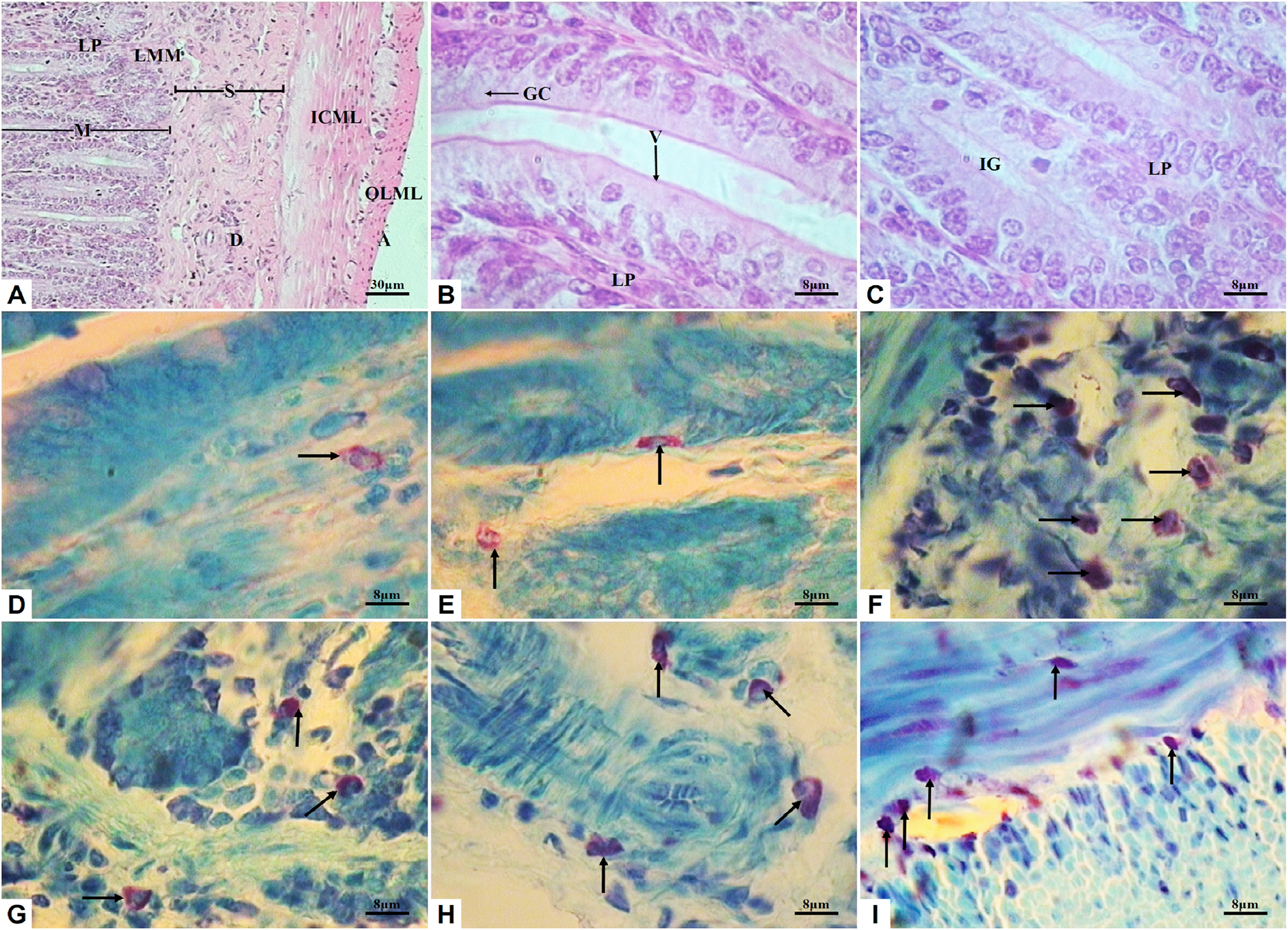
Microstructure and mast cells in the hedgehog’s duodenum (A, magnification of 100X; B to I, magnification of 400X). A to C, HE staining; D to I, toluidine blue staining. Black arrows indicating MCs. M, mucosa; LMM, lamina muscularis mucosa; LP, lamina propria; GC, goblet cell; A, adventitia; ICML, inner circular muscle layer; OLML, outer longitudinal muscle layer; S, submucosa; D, duct; V, villi; IG, intestinal glands.

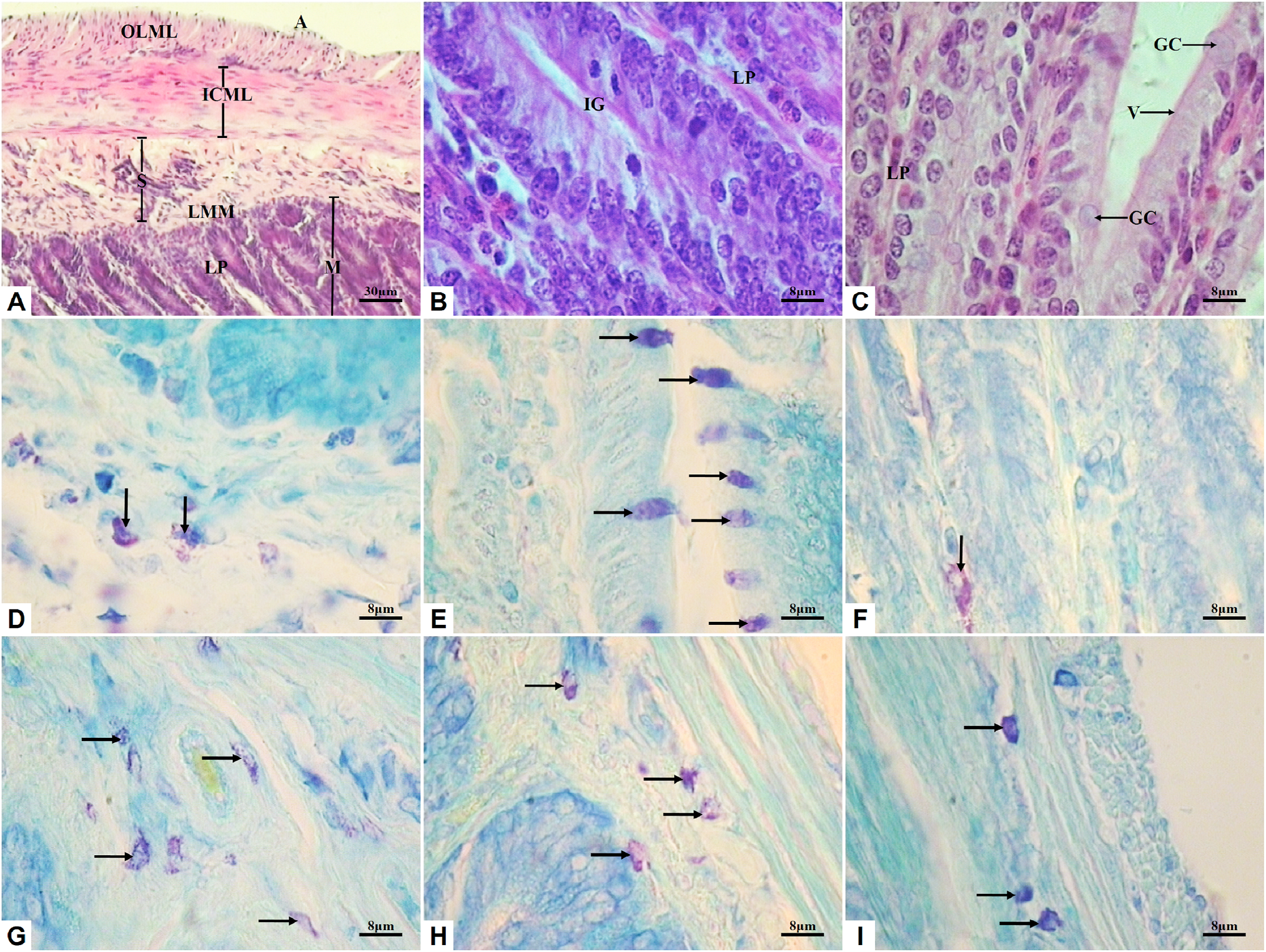
Microstructure and mast cells in the hedgehog’s jejunum (A, magnification of 100X; B to I, magnification of 400X). A to C, HE staining; D to I, toluidine blue staining. Black arrows indicating MCs. M, mucosa; EM, epithelium mucosa; LMM, lamina muscularis mucosa; LP, lamina propria; GC, goblet cell; A, adventitia; ICML, inner circular muscle layer; OLML, outer longitudinal muscle layer; S, submucosa; V, villi; IG, intestinal glands.

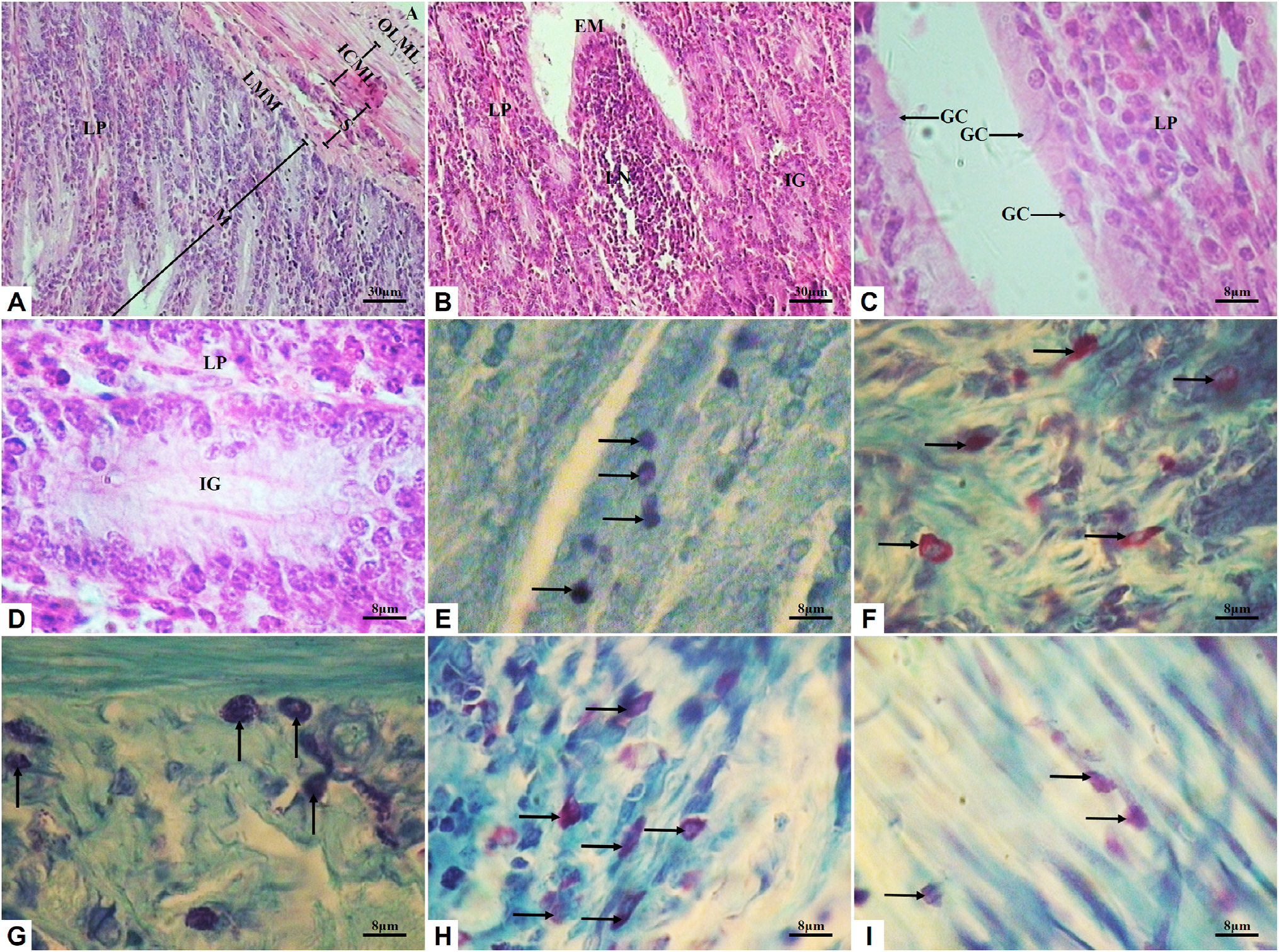
Microstructure and mast cells in the hedgehog’s ileum (A and B, magnification of 100X; C to I, magnification of 400X). A to D, HE staining; E to I, toluidine blue staining. Black arrows indicating MCs. M, mucosa; LMM, lamina muscularis mucosa; LP, lamina propria; GC, goblet cell; A, adventitia; ICML, inner circular muscle layer; OLML, outer longitudinal muscle layer; IG, intestinal glands; LN, lymphoid nodule.


