
Genetic Characterization and Pathological Evaluation of Clade 2.3.4.4b Avian Influenza Virus(H5N8) in Naturally Infected Domestic Ducks in Egyptian Farms


Genetic Characterization and Pathological Evaluation of Clade 2.3.4.4b Avian Influenza Virus(H5N8) in Naturally Infected Domestic Ducks in Egyptian Farms
Sara Magdy Hashim1, Elshaimaa Ismael2, Mohamed Tarek3, Faten Fathy Mohammed1*, Fatma Amer Abdel Reheem3, Rawhia Esawy Doghaim1


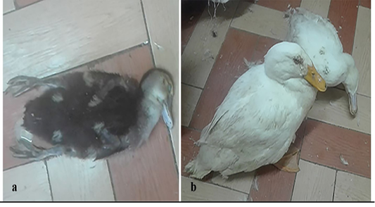
Clinical signs of HPAIV H5N8 infected ducks, (a) Sudani duckling, 60 days old from Giza governorate, showing greenish diarrhea and respiratory distress. (b) Pekin ducks, 120 days old from Giza governorate showing nervous signs (torticollis) including tremor of neck.

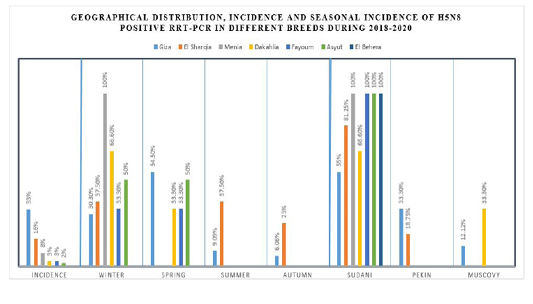
Geographical distribution, incidence and seasonal incidence of H5N8 positive rRt-PCR in different breeds during 2018-2020.
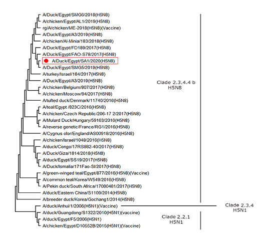
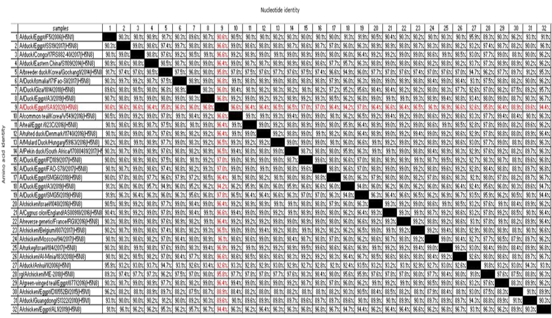
Phylogenetic relationship of HPAI H5N8 local field isolates to other selected AIV isolates based on nucleotide sequence of HA gene. Phylogenetic tree of the HA gene segment of HPAI H5N8 viruses. A phylogenetic tree including a total of 32 HA segments from different H5N8 viruses was obtained using MEGA X software.
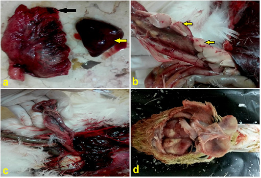
(a) Lung, duck showing congestion and edema (black arrow), spleen showing splenomegaly and congestion (yellow arrow). (b) Pancreas, duck showing necrotic areas (yellow arrows).) with hemorrhage (c) Duodenum, duck showing severe congestion of serosal vasculatures. (d) Brain, duck showing congestion.
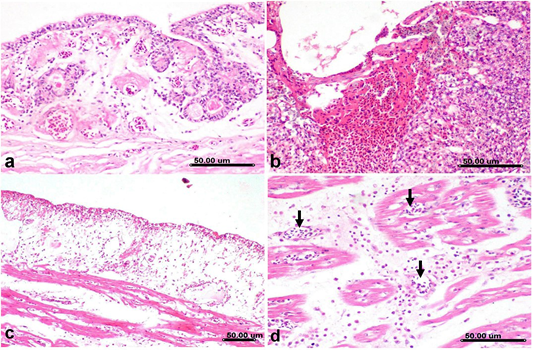
Histological sections of lung and heart from H5N8 infected duck, a) Primary bronchus showing deciliation of lining epithelium with congestion associated with interstitial edema and mononuclear cells infiltration. b) Parabronchus showing hemorrhage with proliferative reaction involving the air capillaries with mononuclear cells infiltration. c) Heart, epicardium showing desquamation of lining mesothelium associated with extensive subepicardial edema, congestion and mononuclear cells infiltration, note the extensive intermuscular edema of cardiac muscle fibers in the vicinity of subepicardial area. d) Myocardium showing necrosis of cardiac myocytes with mononuclear cells infiltration, congestion (arrow) and intermuscular edema. (Stain, H&E).
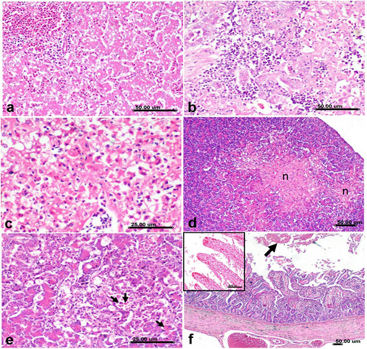
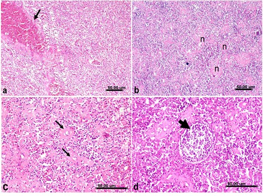
Histological sections of Liver, pancreas and duodenum from H5N8 infected duck, a) Liver showing severe congestion of portal vessels and hepatic sinusoids associated with vacuolization of hepatocellular cytoplasm. b) Liver showing lymphocytic infiltration of hepatic sinusoids. c) Liver showing severe necrotic reaction of hepatocytes with karyorrhexis and pyknosis of their nuclei d) Pancreas showing multifocal necrosis of exocrine pancreatic acini that replaced with eosinophilic necrotic tissue debris (n). e) Pancreas showing vacuolization of pancreatic acinar epithelium associated with apoptosis(arrow). f) Duodenum showing shortening, atrophy and blunting of duodenal villi, severe congestion of submucosal and serosal vessels and mucosal luminal hemorrhage (arrow), the inserted box in upper left corner showing severe ulceration of mucosa with congestion of blood capillaries in lamina propria that was infiltrated by mononuclear cells. (Stain, H&E).

Histological sections of spleen from H5N8 infected duck, showing a) Severe congestion of splenic vessels (arrow) and sinusoids b) Spleen showing severe lymphocytic depletion with marked necrosis of lymphoid follicles (n). c) Fibrinoid necrosis of splenic arterioles(arrow). d) lymphocytic depletion and lymphocytolysis of lymphoid elements comprising the bursal associated follicles (arrow). (Stain, H&E).
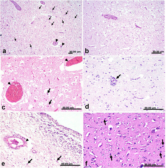
Histological brain sections from H5N8 infected duck showing; a) Congestion and micro- thrombosis of cerebral blood vessels (arrowhead) associated with capillary endothelial proliferation (arrow). b) Spongiosis and vacuolization of cerebral neuropil associate with diffuse gliosis. c) Marked congestion (arrowhead) associated with necrosis of pyramidal neurons involving the cerebellar nucleus(arrow). d) Vasculitis and perivascular lymphocytic cuffing (arrow) associated with gliosis. e) Microthrombosis (arrowhead) and hemorrhage in hippocampus associated with neuronal necrosis (arrow). f) Neuronal degeneration with neuronophagia associated with glisosis of cerebral grey matter. (Stain, H&E)

Histological immunohistochemical sections from H5N8 infected duck. a) Lung showing residence of viral expression in lining epithelium of air capillaries and in infiltrating inflammatory cells. b) Heart showing viral expression in cardiac myocytes. c) Large vessel showing expression of viral antigen in lining endothelium. d) Duodenum showing expression of viral antigen in epithelial lining crypts. e) Spleen showing residence of viral antigen in lymphocytes. f) Brain showing expression of viral antigen in neurons, Capillary endothelium and glia cells.


