Effects of Feed Additives on Chicken Growth and Their Residues in Meat Instigating Deleterious Consequences on the Liver Health of Consumers - A Prospective Human Study
Effects of Feed Additives on Chicken Growth and Their Residues in Meat Instigating Deleterious Consequences on the Liver Health of Consumers - A Prospective Human Study
Saara Ahmad1,*, Iftikhar Ahmed2, Saida Haider3, Zehra Batool4, Laraib Liaquat3, Fatima Ahmed5, Asra Khan1, Tahira Perveen3, Mirza Jawad ul Hasnain6, Saima Khaliq7 and Saad Bilal Ahmed8
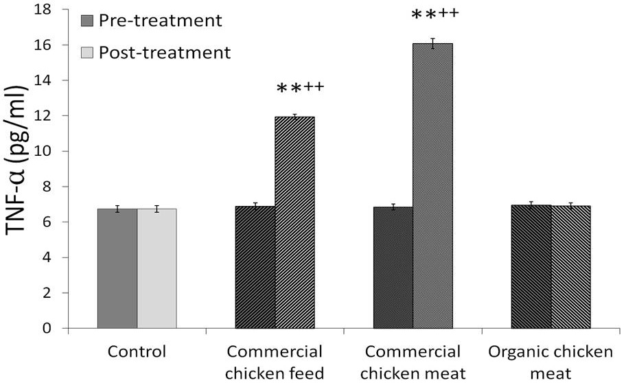
Effects of six weeks administration of commercial chicken feed, meat and organic meat on the levels of THF-alpha. Levels were estimated before and after the experiment. Values are mean ± SEM (n=25). **p<0.05 from respective group A rats; ++p<0.01 from pre-treatment values.
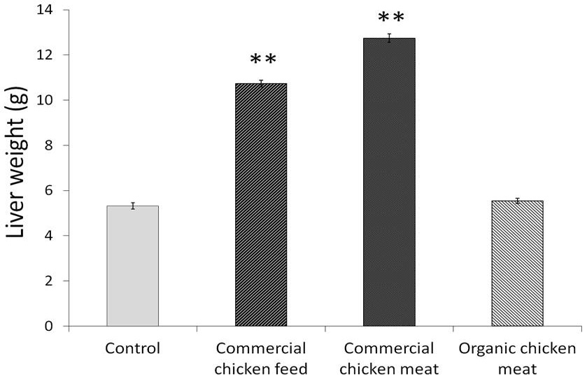
Liver weight of animals following the administration of commercial chicken feed, meat and organic chicken meat. Values are mean ± SEM (n=25). **p<0.05 with respect to group A.
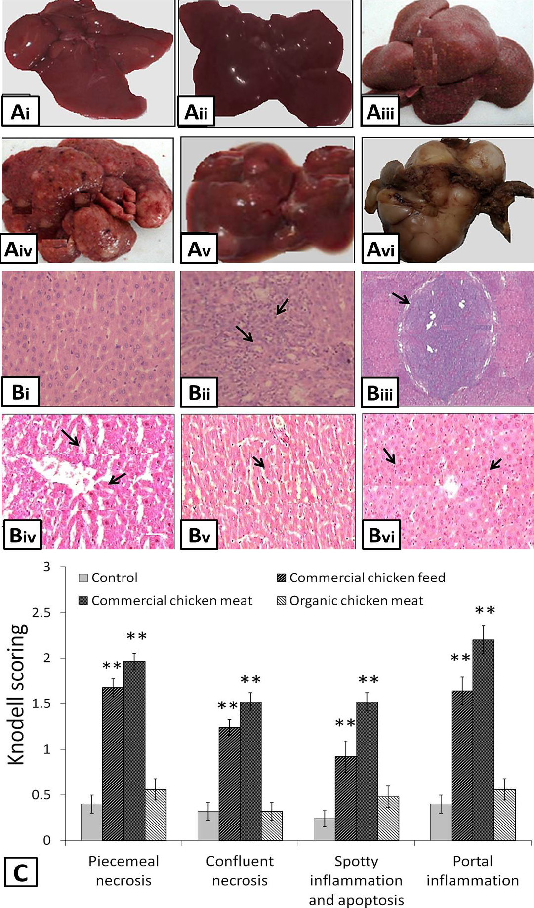
A, morphology of liver observed after the intake of commercial chicken feed, meat and organic meat. (i) and (ii) showed normal morphology of liver isolated from control and organic meat fed animals, (iii) inflamed, (iv) inflamed and nodular, (v) nodular and (vi) cirrhotic liver were seen in animals fed on commercial chicken feed and meat for six weeks. B, histopathological examination of liver samples showing the presence of (i) normal cells in control group A rats and organic chicken meat fed animals, (ii) Inflamed cells, (iii) cirrhotic nodule (iv) portal inflammation (v) confluent necrosis and (vi) spotty inflammation, necrosis with apoptosis were observed in liver cells isolated from the animals fed on commercial chicken feed and meat. C, Knodell scoring was done to assess the portal inflammation, necrosis, apoptosis and cirrhosis in liver samples. Values are mean ± SEM (n=25). **p<0.05 with respect to controls.


