
Effect of Estradiol Benzoate on Epididymis Duct in Male Wistar Ratsat Pre-Puberty


Effect of Estradiol Benzoate on Epididymis Duct in Male Wistar Ratsat Pre-Puberty
Manar Mousa Alhussein*, Eman Faisal Albghdady
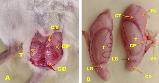
(A) Ventral view of mature wistar rats aged 66 days showed male genital system (B) Showed epididymis duct lie along dorsal surface of testis (T) and epididymal segments: caput (CT), corpus (CP), cauda (CD) that connect with testes by ligament (LG) and continuous with vas deference (VD).
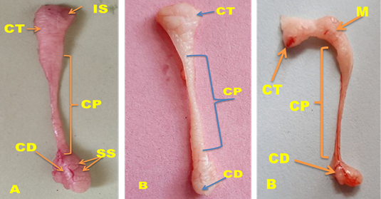
Gross anatomy of the epididymis segments: caput (CT), corpus (CP), cauda (CD) of wistar Rats aged 66 days that split into lobules or sub-segmental subdivision (SS). (A) Epididymis duct non-treated group (G1). (B) Epididymis duct in treated group (G2) with 50µg/rat of estradiol benzoate that was appeared irregular in shape as malformed (M) (right picture).
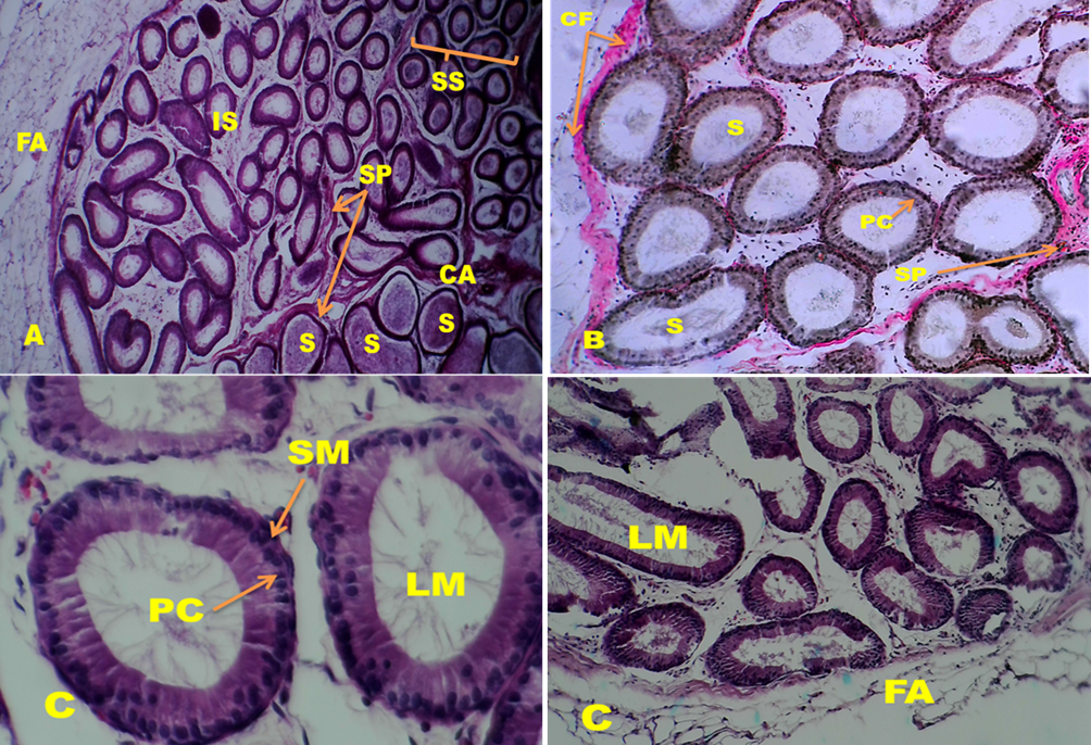
Microscopic section of epididymis segments (A) Initial (IS) and caput segments (CA) of Wistar Rats aged 66 days in non-treated groups showed were surrounded by friable amount of fat (FA) and divided into sub-segmental division (SS), by loose connective tissue septa (SP) rich with collagen fibers (CF). Principle cells (PC) one type of epithelial cells lined tubules that full with sperms. (A) HandE 40X; (B) Initial (IS), of non-treated groups (G1) showed ducts surrounded by clear quantity of collagen fiber(CF) and ducts divided by loose connective tissue septa (SP), each ducts full with sperm(S)Van Gieson’s stain 100X(C) Initial segments of treated groups(G2) showed lumen duct (LM) devoid of sperm, each tubule surrounded from out by layers of smooth muscle (SM), and lined by epithelia that contain most cell such as principle cell(PC). (C)HandE 400X (left picture), 100X (right picture).


Transverses section of corpus of epididymis of Wistar Rats aged 66 days. (A) corpus in non-treated group (G1) showed tubulars lined with pseudostratified columnar epithelial that consist of principle cell (PC) and it was full with sperms (S). HandE, 100X (B) Corpus of epididymis of treated groups (G2) with 50µg/rat of estradiol benzoate, showed tubules separated by loose connective tissue (CO) as septa (SP) that rich with of collagen fibers (CA) and surrounded by smooth muscle fibers (SM). Tubular lumen (LM) devoid of sperms and there was hyperplasia (HP) with increased in thickness of epithelial and appeared cribriform forms (CR), and increase of bubbly lipid vacuolization (V) in the supra-nuclear area of the principal cells and inflammatory infiltrate (FC). (B) Left: HandE, 200X; Right PAS Stain 100X.

Transverses section of cauda of epididymis of Wistar Rats aged 66 days (A) cauda in non-treated groups(G1) showed pseudostratified columnar epithelial that consist of principle cell(PC), basal cell (BC), lined tubules that full with sperms (S) and between tubules loose connective tissue (CO). HandE, 200X. (B) Cauda of epididymis of treated groups (G2) with 50µg/rat of estradiol benzoate showed most tubules lumen (LM) irregular in shape, shrinking, smaller and devoid of sperms or had debris (DS). There was hyperplasia (HP) with increased in thickness of epithelial and appeared cribriform forms (CR) and there is large amount of collagen fibers (CF) between tubules. Van Gieson’s stains 200X.

