Effect of Gluten Containing Diet on Pristane Induced Lupus Prone Mice
Effect of Gluten Containing Diet on Pristane Induced Lupus Prone Mice
Muhammad Mansoor, Zaigham Abbas and Nageen Husssain*
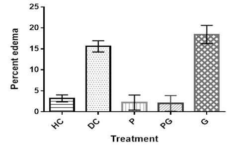
Paw edema (%) after treatment in pristane induced lupus mice.
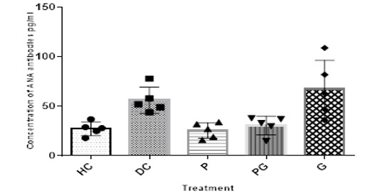
ANA autoantibodies level in pristane induced lupus mice after treatment.
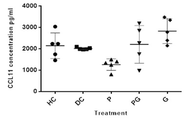
CCL11 concentration in pristane injected mice after treatment.
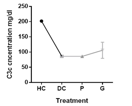
C3c level in pristane induced mice after treatment.
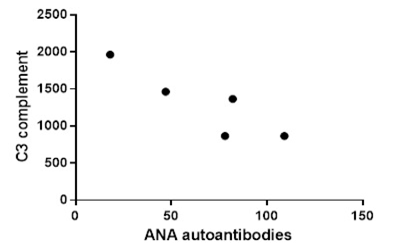
Correlation between ANA and C3 level in pristane induced lupus mice.
Histological structure of kidney of positive control mouse. Thich black arrows in (x100) showing epithelial crescent squashing the glomerular tufts from all sides, yellow arrow showing necrocitising glomerulus, black arrow in (x400) showing segmental glomerular sclerosis.
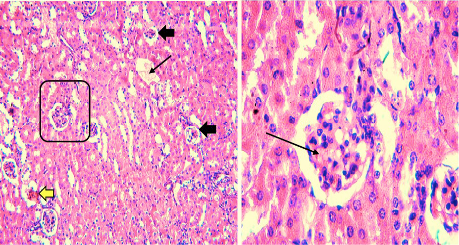
Histological structure of kidney of prednisone treated mouse. Black arrows in (x100) showing affected glomeruli. Not a single glomerulus was normal showing severe lupus nephritis. Thick black arrow in (x400) showing hyaline arteriosclerosis and thin arrow showing glomerular necrosis, brown arrow showing immune deposits of membranous nephropathy, yellow arrow showing tubules with neutrophils.
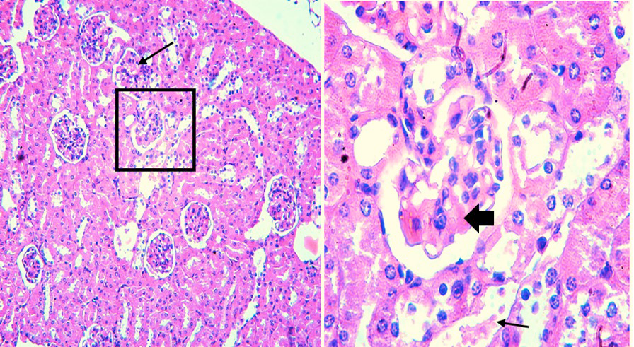
Histological structure of kidney of mouse fed on prednisone + gluten containing diet. Arrow in (x100) showing glomerulonephritis. Most of other glomerulus in (x100) are normal. Thick black arrow showing partial hyaline sclerosis and thin arrow showing tubules with neutrophils.
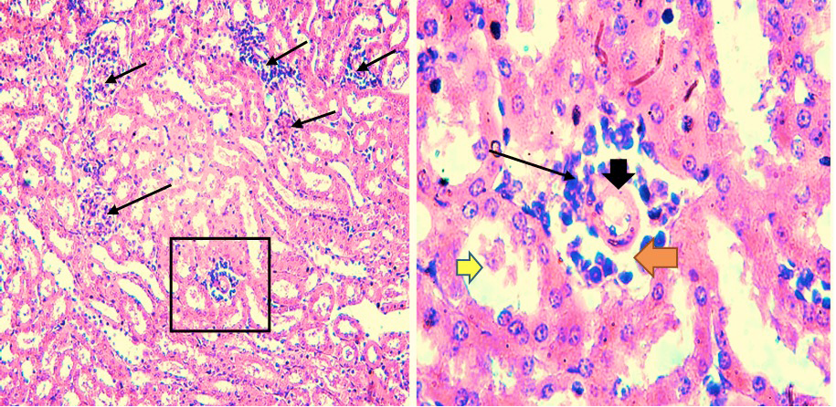
Histological structure of kidney of mouse fed on gluten containing diet. Black arrows in (x100) and yellow arrow in (x400) showing rapidly progressive glomerulonephritis, red arrow showing tubules with neutrophils and bl


