miR-216b Downregulates Intestinal IL2RB to Inhibit the Invasion of Echinococcus granulosus Eggs in the Kazakh Sheep
miR-216b Downregulates Intestinal IL2RB to Inhibit the Invasion of Echinococcus granulosus Eggs in the Kazakh Sheep
Xuhai Wang1,2, Xin Li1, Fangyuan Yuan1, Chaocheng Li1, Bin Jia1* and Song Jiang1*
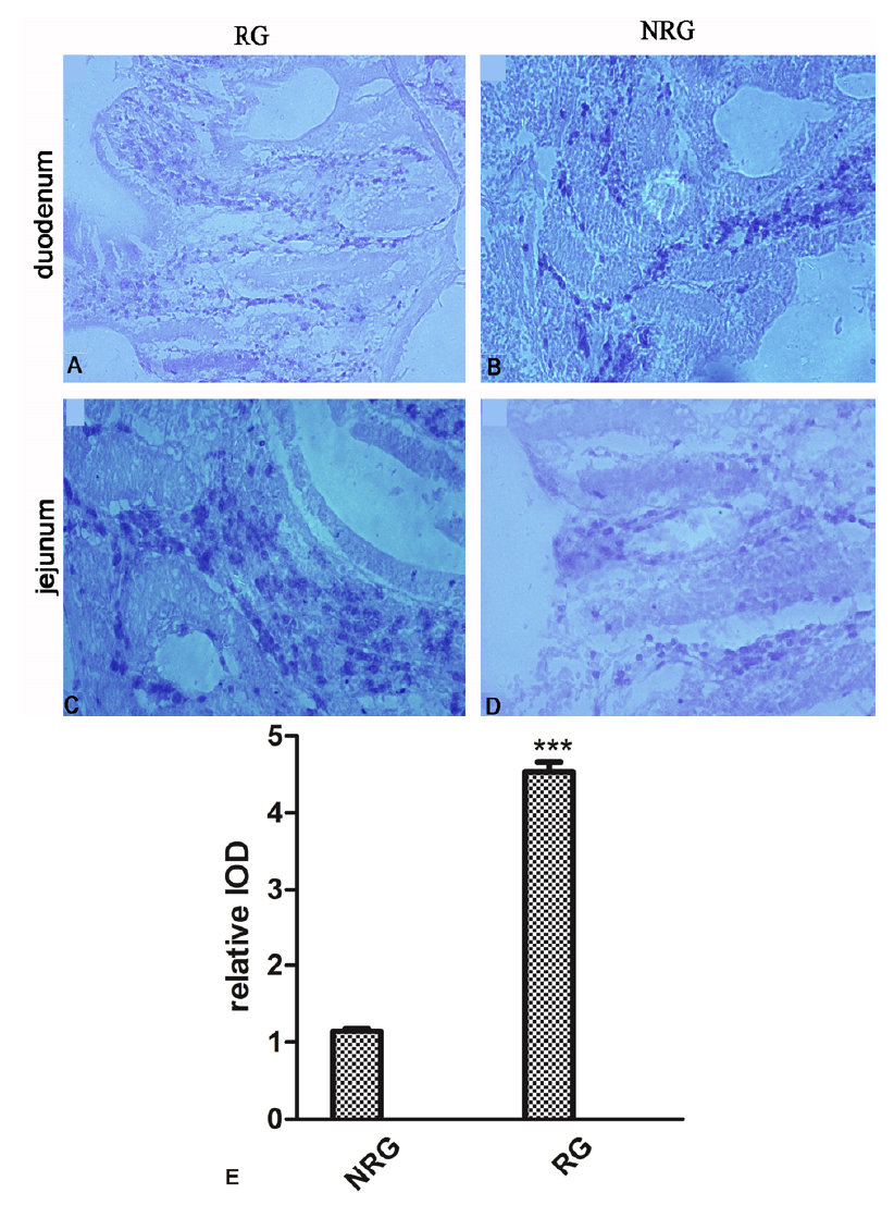
The distribution of IL2RB in the small intestine of sheep. Intestinal IL2RB expression in NRG and RG sheep was determined by immunohistochemical staining. A, C: Strong IL2RB positive expression was observed in T cells of mucosal layer and musclar layer. B, D: The positive signal of IL2RB was only detected in mucosal layer, especially in the lamina propria and epithelium. E: The average IOD was obtained by analysing IL2RB IHC in four random felds of each slide. Upper panel: representative image; lower panel: quantitative analysis. Results are presented as the mean± SEM (magnifcation, 200×, scale bars= 100μm).
miR-216b and IL2RB protein levels are inversely correlated in RG and NRG sheep intestinal tissues. miRNA in situ hybridization for miR-216b was performed on intestinal sections from RG and NRG controls (green, miR-216b; blue, DAPI nuclear staining). (Magnifcation 200×, scale bar= 200μm).
The IL2RB 3’-UTR contains conserved miR-216b target sites. A: The fold-changes of seven miRNAs predicted by three algorithms in RG intestinal tissues compared to NRG (control), as analysed by high-throughput sequencing. B: Schematic illustration of conserved duplexes formed by IL2RB 3’UTR and miR-216b interactions. The predicted free energy of the hybrid is noted. Paired bases are marked by a blue line. The complementary seed sites are marked in blue. C: Quantitative real-time PCR analysis of IL2RB expression in 293T cells transfected with miR-216b mimics, miR-216b inhibitor, IL2RB-W or IL2RB-M. Data are presented as the mean±SEM from three independent experiments (293T group: ***p=0.000092, IL2RB-M vs. IL2RB-W). D: Relative luminescence intensity detected by a Hamamatsu optical analyze reader after miR-216b mimics and dual-luciferase vectors were co-transfected into 293T cells (*** P<0.01).


