Differential Regulation of Hsp70 Expression in Six Lizard Species under Normal and High Environmental Temperatures
Differential Regulation of Hsp70 Expression in Six Lizard Species under Normal and High Environmental Temperatures
Wei Dang1, Ning Xu1, Wen Zhang1, Jing Gao2, Handong Fan1 and Hongliang Lu 1,*
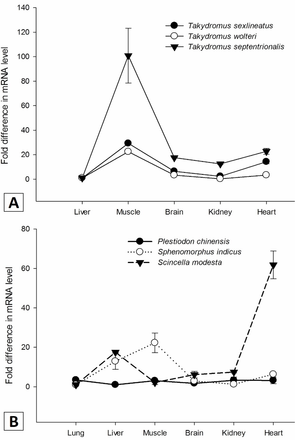
Hsp70 expression in different tissues of the six lizards species detected by quantitative real time reverse transcriptase PCR. Hsp70 expression levels in different organs were normalized to that of β-actin mRNA. For Takydromus, the normalized Hsp70 mRNA level in liver was set as 1. For P. chinensis, Sp. indicus and Sc. modesta, the normalized Hsp70 mRNA level in lung was set as 1. Vertical bars represent means±SE (n=5).
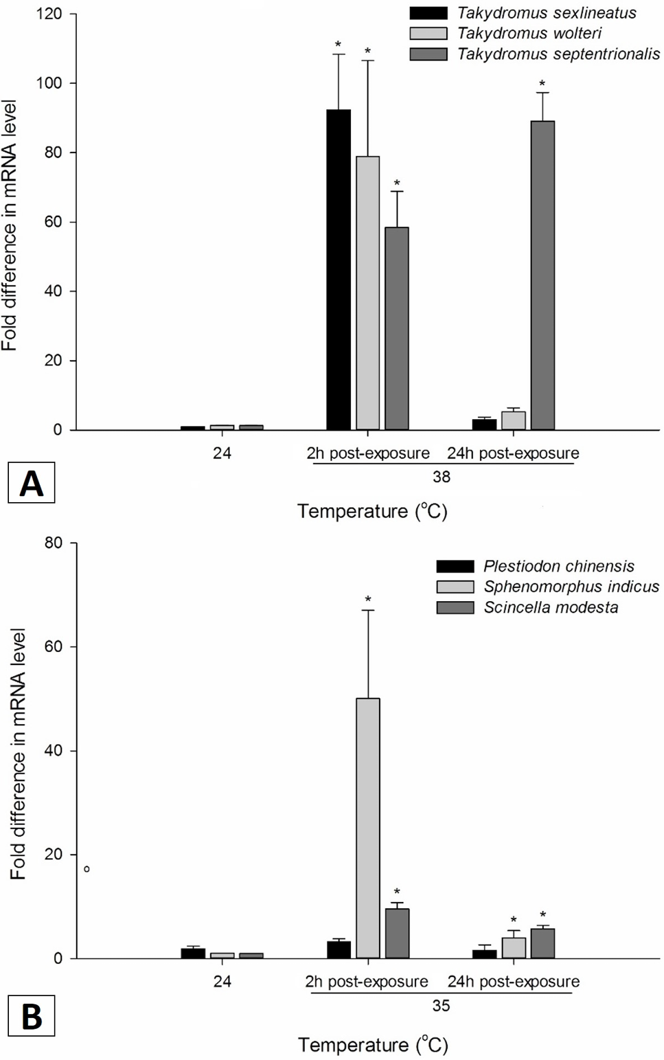
Hsp70 Expression of six lizards species in response to heat shock. Hsp70 expression in liver was determined by quantitative real time reverse transcriptase PCR at various times post-exposure. The mRNA level of Hsp70 was normalized to that of β-actin mRNA. Values are shown as means ± SE (n = 5). Significances between room temperature lizards and heat post-exposure lizards are indicated with asterisks. *P < 0.05.
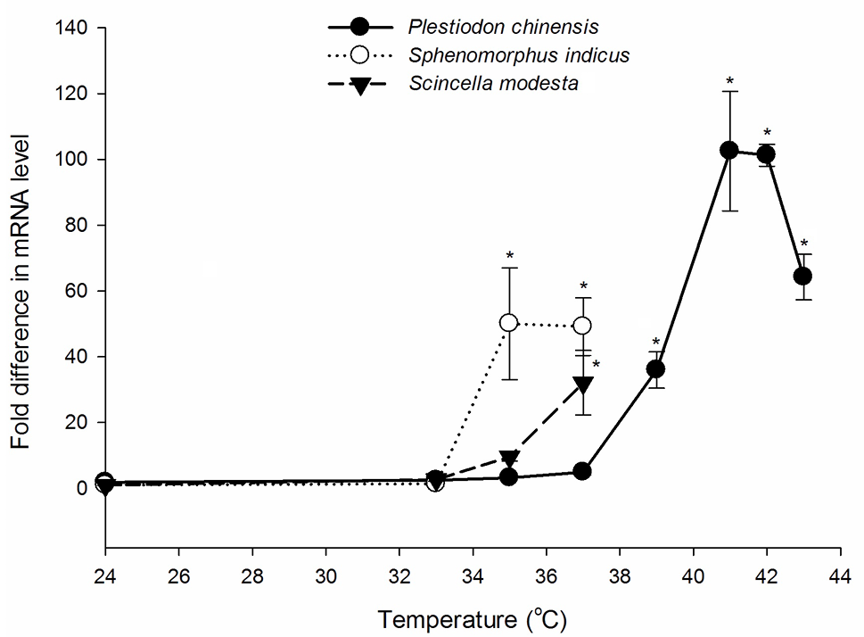
Hsp70 expression of P. chinensis, Sp. indicus and Sc. modesta in response to different temperature in short time. Hsp70 expression in liver was determined by quantitative real time reverse transcriptase PCR at various times post-exposure. The mRNA level of Hsp70 was normalized to that of β-actin mRNA. Values are shown as means ± SE (n = 5). Significances between room temperature lizards and heat post-exposure lizards are indicated with asterisks. *P < 0.05.


