
Development of a Test System Based on Recombinant GM6 Antigen from Trypanosoma evansi for the Determination of Surra in Horses


Development of a Test System Based on Recombinant GM6 Antigen from Trypanosoma evansi for the Determination of Surra in Horses
Nurlan Akhmetsadykov1*, Tanatar Kydyrov2, Moldir Akhmetzhanova1, Gulnazi Akhmetova3, Maxat Berdikulov4


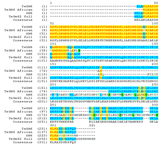
Comparative analysis of trypanosomal antigen amino acid sequences isolated from T. evansi across various regions of Kazakhstan
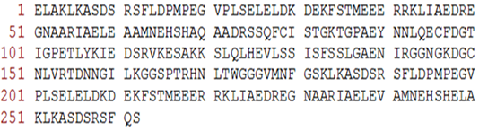
The amino acid sequence of GM6 isolated from T. evansi

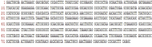
The nucleotide sequence of GM6 trypanosomal antigen gene isolated from T. evansi

PCR results of E. coli clones transformed with pET28/GM6 carrying a trypanosomal antigen insert
Note: 1 – DNA markers; 2-19 – E. coli clones transformed with pET28/GM6.
Determination of trypanosomal protein expression depending on time of incubation in IPTG
Note: 1 – cell lysate; 2 – supernatant after centrifugation of cell lysate; 3 – pellet after centrifugation of cell lysate; 4 – pellet of cell lysate without IPTG; 5 – cell lysate pellet after 6 hours’ incubation with IPTG; 6 – pellet of cell lysate after 18 hours’ incubation with IPTG; mm – molecular weights marker.
Electrophoregram of purified preparations of trypanosomal antigen GM6
Note: 1-4 – Purified fractions of trypanosomal antigen; М – molecular weights marker.

Immunoblotting of recombinant trypanosomal antigen GM6 T. evansi
Note: A – antibodies against 6-His; B – serum from trypanosomosis infected horse; 1 – recombinant GM6 protein prior to expression; 2 – recombinant GM6 protein after expression; M – molecular weights marker.

Immunoenzymatic analysis of recombinant trypanosomal antigen with sera from diseased animals
Note: H1-H4 – serum of a horse experimentally infected with T. evansi trypanosomosis; H5-H8 – serum of a horse experimentally infected with T. equiperdum trypanosomosis; H9, H10 – serum of horses suffering naturally from trypanosomosis; D1-D5 – sera of donkeys suffering naturally from trypanosomosis.
Immunoenzymatic analysis of recombinant trypanosomal antigen with sera from healthy animals
Note: H11-18 – healthy horse serum; D7-15 – healthy donkey serum.


