
Detection of Non-Cytopathic Enteroviruses in Supernatant of RD and L20B Cell Cultures


Detection of Non-Cytopathic Enteroviruses in Supernatant of RD and L20B Cell Cultures
Adewumi Moses Olubusuyi1, Ogunsakin Temitope Rachael1, Ogunrombi Sefiu Babatunde1, Ojeamiren Iduda1, Olawole Alaba Samuel1, Faleye Temitope Oluwasegun Cephas1,2* and Adeniji Johnson Adekunle1,3

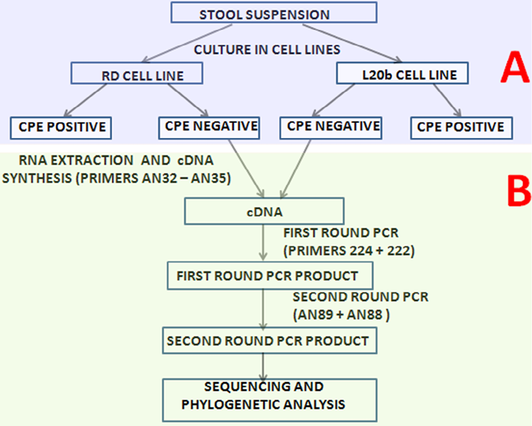
The algorithm followed in this study. The section depicted ‘A’ was executed by the WHO polio laboratory in Ibadan, Nigeria while the section depicted ‘B’ was executed in this study. The cell culture supernatants from RD and L20B cell culture tubes that showed no CPE were collected from the WHO polio laboratory and analyzed in this study.

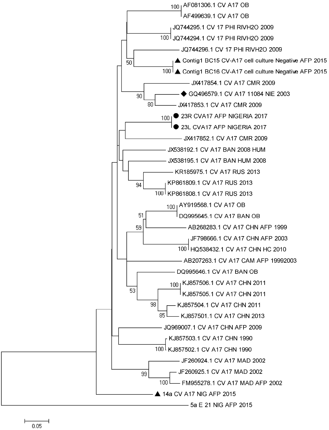
Phylogenetic tree of genetic relationship between VP1 nucleotide sequences of CV-A17. The phylogenetic tree is based on an alignment of the partial VP1 sequences. The newly sequenced strains are indicated with black circle. Strains detected in Nigeria in 2003 and 2015 are indicated with black diamond and triangle, respectively. Bootstrap values are indicated if >50%.

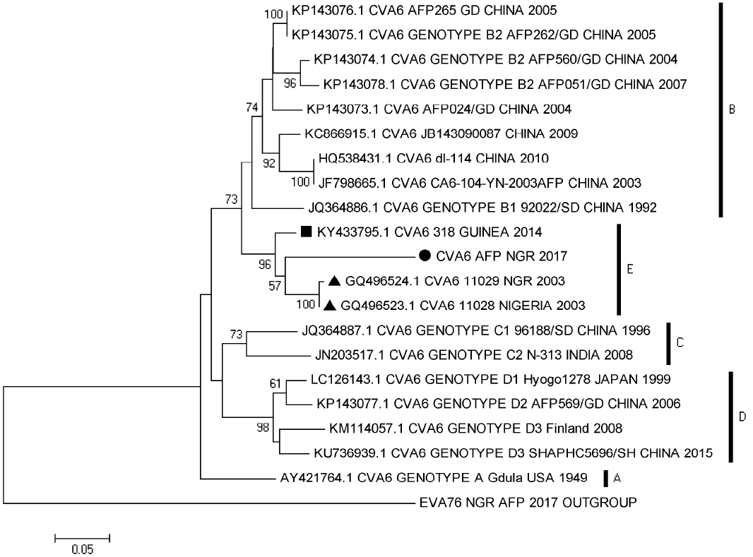
Phylogenetic tree of genetic relationship between VP1 nucleotide sequences of CV-A6. The phylogenetic tree is based on an alignment of the partial VP1 sequences. The newly sequenced strains are indicated with black circle. Strains detected in Nigeria in 2003 are indicated with black triangle, while that detected in Guinea in 2014 is indicated with a black square. Bootstrap values are indicated if >50%.

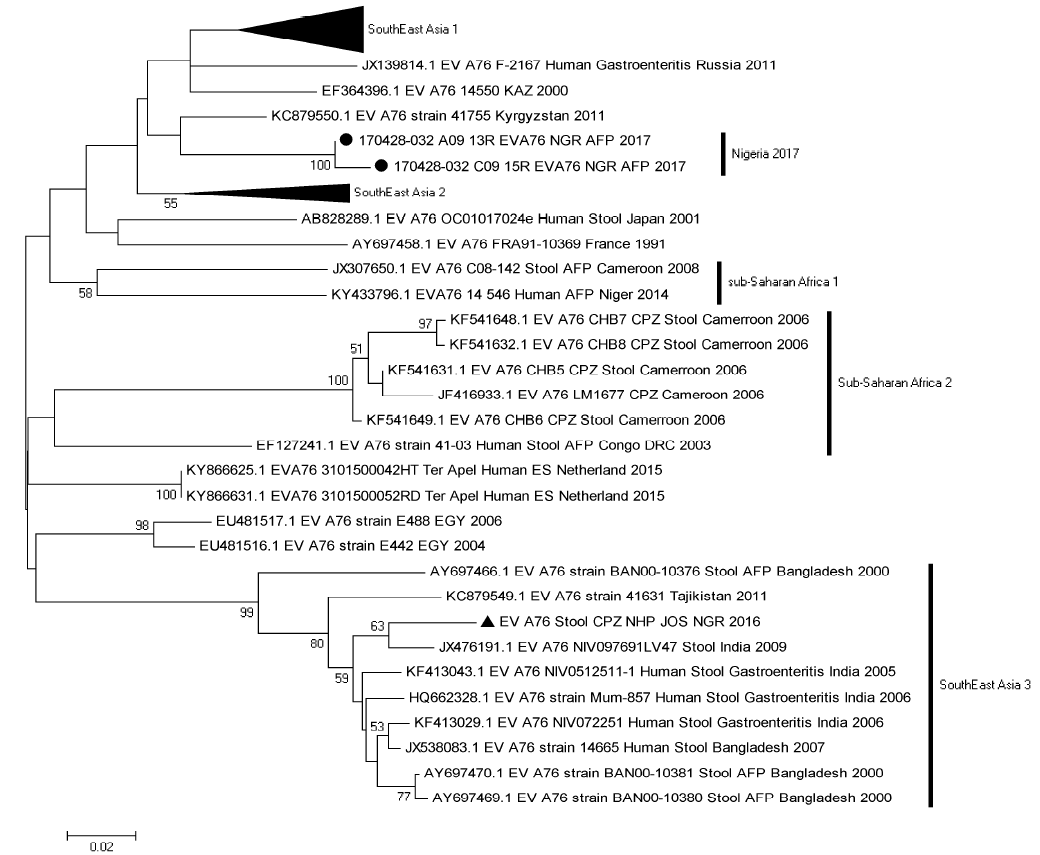
Phylogenetic tree of genetic relationship between VP1 nucleotide sequences of EV-A76. The phylogenetic tree is based on an alignment of the partial VP1 sequences. The newly sequenced strains are indicated with black circle. A strain detected in a nonhuman primate in Nigeria in 2016 is indicated with black triangle. Bootstrap values are indicated if >50%.

