
Dermatophytosis in Cats: Clinical Signs and Identification of Etiological Agent


Dermatophytosis in Cats: Clinical Signs and Identification of Etiological Agent
Alsi Dara Paryuni1, Soedarmanto Indarjulianto1, Tri Untari2, Sitarina Widyarini3*

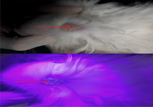
Figure 1:
Lesions from dermatophyte infection in body part of cat with crust and alopecia in the skin (red arrow); Skin lesions of cat with fluorescence (apple blue-green color) under Wood’s lamp examination (red arrow).

Figure 2:
Fungal colony of M. canis in DTM (A); M. canis macroconidia (B).
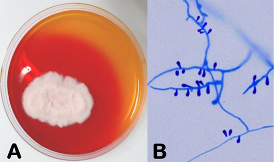
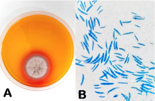
Figure 3:
Fungal colony of T. mentagrophytes in DTM (A); T. mentagrophytes microconidia (B).

April 2023
Vol. 11, Iss. 4, Pages 517-694

