Construction and Protective Efficacy of Egy-H5 DNA Vaccine from Local Egyptian strain H5N1 using Codon Optimized HA gene
Construction and Protective Efficacy of Egy-H5 DNA Vaccine from Local Egyptian strain H5N1 using Codon Optimized HA gene
Wesam Hasan Mohamed Mady1*, Bing Liu2, Dong Huang2, Abdel Satar Arafa1, Mohamed Khalifa Hassan1, Mona Mehrez Aly1, Pucheng Chen2, Yongping Jiang2* and Hualan Chen2
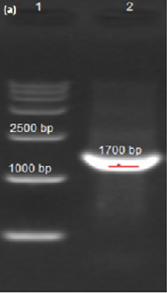
Agarose Gel electrophoresis showing the screening of positive clones, Lane 1 is the molecular weight marker and lane 2 is the inserted opti-HA at the expected weight 1700 bp.
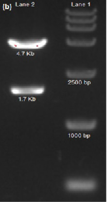
Agarose gel electrophoresis showing the pattern of the digestion analysis. Lane 1 is the molecular weight marker and Lane 2 is the double digested pCA-Egy-H5 plasmid DNA showing 2 bands, the upper band is the pCAGGS plasmid at 4.7 Kb and the Lower band is the opti HA at 1.7 Kb.
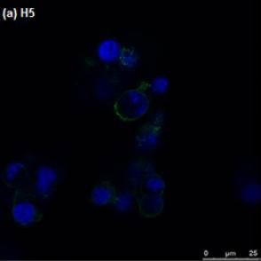
The 293T HEK cells transfected with pCA-Egy-H5 showing the bright specific fluorescence.
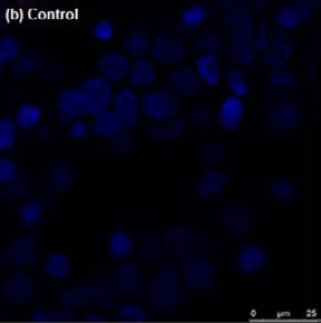
The negative Control 293T HEK cells not transfected did not show any fluorescence.
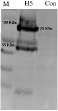
Showing the western blotting of the expressed H5 protein in 293T HEK cells, M is the molecular weight marker, H5 is the expressed HA protein showing the specific band at 70 KDa, and Con = the cell negative control not transfected.
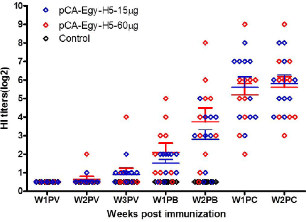
The HI antibody titers induced by pCA-Egy-H5 post immunization. W1PV: 1-week post vaccination; W2PV: 2 weeks post vaccination; W3PV: 3 weeks post vaccination; W1PB: 1-week post boost; W2PB: 2 weeks post boost; W1PC: 1-week post challenge; W2PC: 2 weeks post challenge.


