

Consequence of Exogenous Administration of Oxytocin on Reproductive and Productive Parameters during Postpartum Involution Period in Newly Calved Nili-Ravi Buffaloes


Consequence of Exogenous Administration of Oxytocin on Reproductive and Productive Parameters during Postpartum Involution Period in Newly Calved Nili-Ravi Buffaloes
Saeed Murtaza1, Abdul Sattar1*, Nasim Ahmad1, Muhammad Ijaz2, Maqsood Akhtar3 and Muhammad Shahzad4

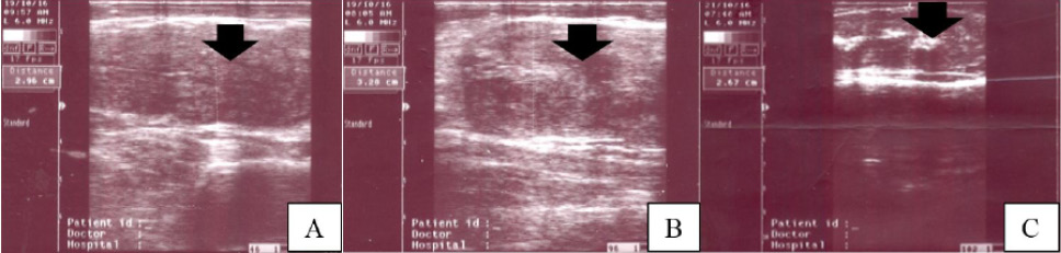
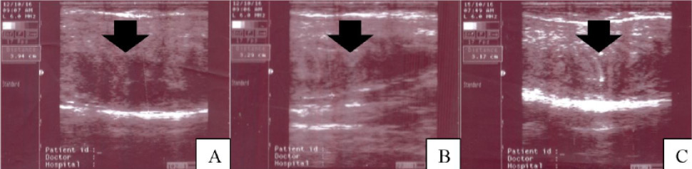
Ultrasonographs of cervix among treatments: Control; A, Low dose 10IU; B, High dose 30IU; C at fifth week near complete involution. Arrow heads showing the longitudinal ultrasound images of cervix in three treatments: A, B and C with enhanced echogenicity in C part of Figure 1.
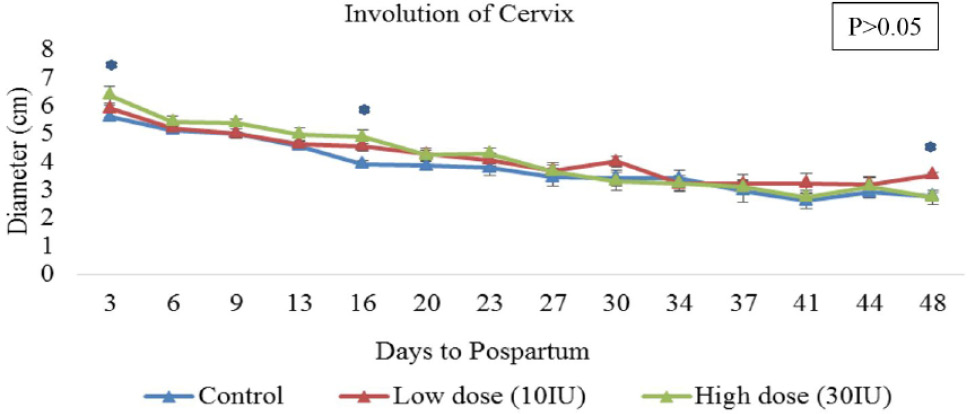
Comparison of means ± S.E (cm) of cervical diameter among three treatment groups between 3 to 48 days of postpartum period in Nili-Ravi buffaloes; Means± S.E with asterisk sign on days: 3; 16 and 48 of postpartum differs among treatment groups at P < 0.05.
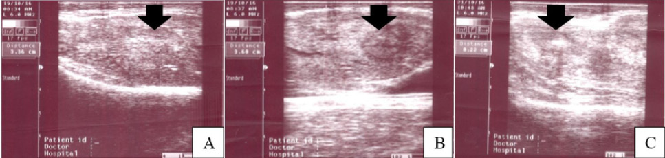
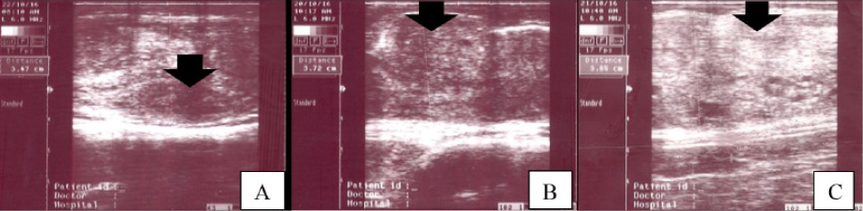
Ultrasonographs of body of uterus at involution in three treatment groups: Control; A, Low dose 10IU; B, High dose 30IU; C at fifth week near complete involution. Arrow heads in A and B parts of Figure 3 indicate normal echogenicity while hyperechoic spots in C part of Figure 3.
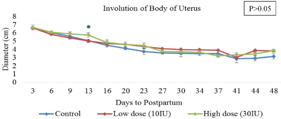
Comparison of means ± S.E (cm) of the diameter of body of uterus among three treatment groups between 3 to 48 days of postpartum period in Nili-Ravi buffaloes; Means± S.E with asterisk sign on day 13 of postpartum differs among three treatment groups at P < 0.05.

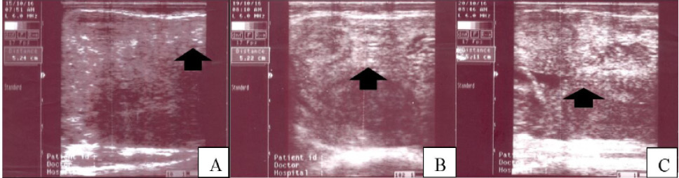
Ultrasonographs of gravid horn in three treatment groups: Control; A, Low dose 10IU; B, High dose 30IU; C at fifth week near complete involution. Cross sectional ultrasound images of three treatment groups in Figure 5 showing normal echogenicity in A and B parts while enhanced echogenicity in C part.
Ultrasonographs of non-gravid horn in three treatment groups: Control; A, Low dose 10IU; B, High dose 30IU; C at fifth week near complete involution. Arrow head in C part of Figure 7 shows more echogenicity parallel to A and B parts.
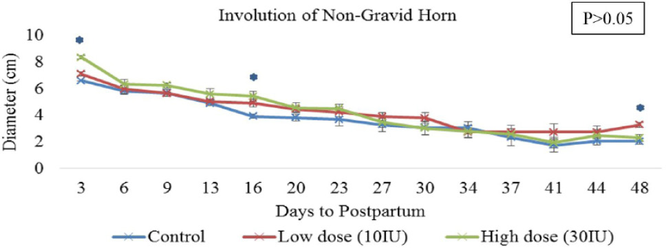
Comparison of means ± S.E (cm) of non-gravid horn diameter among three treatment groups between 3 to 48 days of postpartum period in Nili-Ravi buffaloes. Means± S.E with asterisk sign on days: 3, 16 and 48 of postpartum differs among three treatments at P < 0.05.

Ultrasonographs of caruncles at involution in three treatment groups: Control; A, Low dose 10IU; B, High dose 30IU; C at the end of second week near complete regression. Arrow heads show caruncular points with enhanced echogenicity in C part of Figure 9.
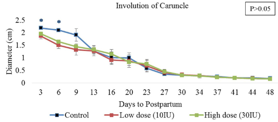
Comparison of means ± S.E (cm) of caruncular diameter among three treatment groups between 3 to 48 days of postpartum period in Nili-Ravi buffaloes. Means± S.E with asterisk sign on days; 3 and 6 of postpartum differs at P < 0.05 among three treatment groups.
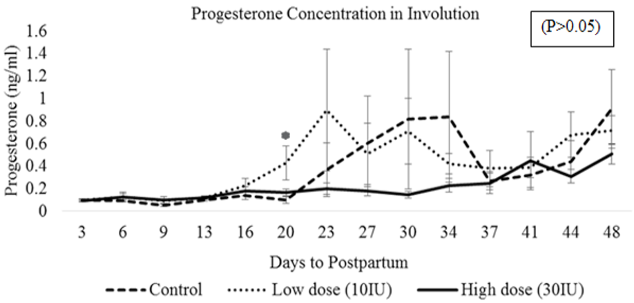
Comparison of means ± S.E (cm) of P4 concentration among three treatment groups between 3 to 48 days of postpartum period in Nili-Ravi buffaloes. Means± S.E with asterisk sign on day 20 of postpartum differs among three treatments at P < 0.05.

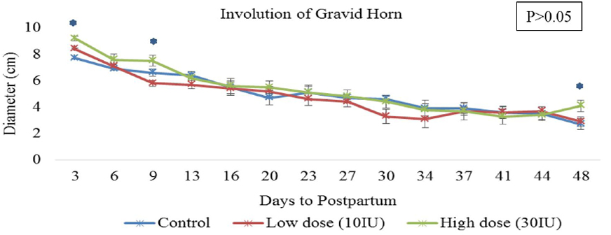
Comparison of means ± S.E (cm) of gravid horn diameter among three treatment groups between 3 to 48 days of postpartum period in Nili-Ravi buffaloes. Means± S.E with asterisk sign on days: 3, 9 and 48 of postpartum differs at P < 0.05 among three treatment groups.


