

Comparison of Cranium Shape in Hamdani and Awassi Sheep using Dorsal and Lateral Landmarks


Comparison of Cranium Shape in Hamdani and Awassi Sheep using Dorsal and Lateral Landmarks
Ismail Demircioglu1, Yasin Demiraslan2, Barıs Can Güzel3*, Ali Koçyigit4 and Aysegül Demircioglu5
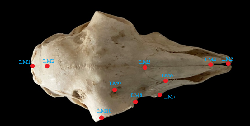
Dorsal landmarks: LM1, External occipital protuberance; LM2, Junction of sutura coronalis and sutura interfrontalis; LM3, Junction of sutura interfrontalis; sutura internasalis and frontonasal suture; LM4, Anterior edge of sutura internasalis; LM5, Anterior edge of fi ssura interincisiva; LM6, Fissura nasomaxillaris; LM7, Tuber faciale; LM8, Medial angle of orbita; LM9, Foramen supraorbitale, LM10; Posterio-ventral corner of margo supraorbitalis.

Lateral landmarks: LM1, Anterior edge of incisiv bone; LM2, Infraorbital foramen; LM3, Anterio-dorsal edge of PM1; LM4, Caudal edge of M3; LM5, Middle point of zygomatic arch; LM6, External acoustic pore; LM7, Ventral edge of jugular process; LM8, External occipital protuberance; LM9, Ventral edge of occipital condyle; LM10, Middle point of margo supraorbitalis; LM11, Medial angle of orbita; LM12, Fissura nasomaxillaris; LM13, Anterior edge of septal process.
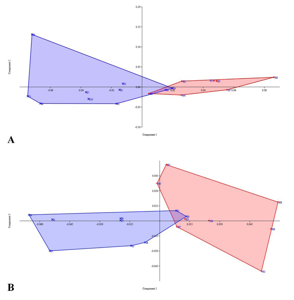
Distribution of individuals on the graph based on the first principal component; A, Dorsal; B. Lateral; Red, Hamdani, Blue, İvesi.

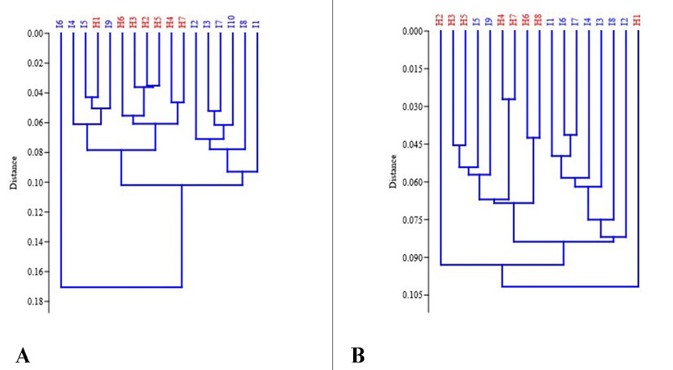
Graphical representation of hierarchical closeness of individuals. A, Dorsal; B, Lateral, Red, Hamdani; Blue, İvesi.

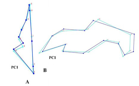
Wireframe graphical view of shape differences according to the first principal component A. Dorsal, B. Lateral, dark blue color represents the average shape based on the primary principal component.


