Comparative Study on Induction of Apoptosis in A549 Human Lung Adenocarcinoma Cells by C2 Ceramide or Ceranib-2
Comparative Study on Induction of Apoptosis in A549 Human Lung Adenocarcinoma Cells by C2 Ceramide or Ceranib-2
Gökhan Kuş1,*, Hatice Mehtap Kutlu2, Djanan Vejselova2 and Emre Comlekci3
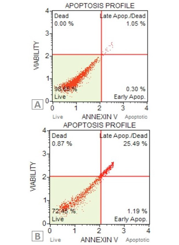
Apoptosis quantification of A549 cells treated with C2 ceramide for 24 h. A, Untreated A549 cells; B, C2 Ceramide treated A549 cells.
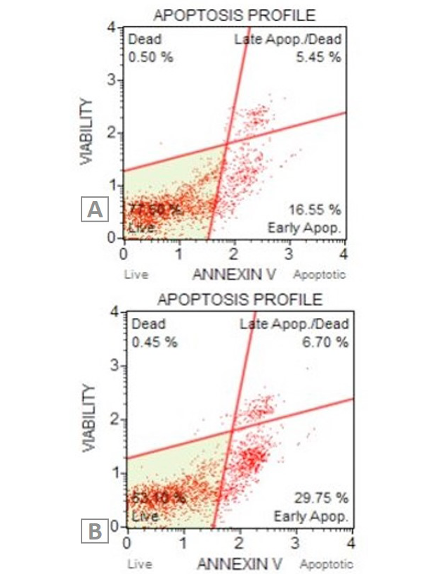
Apoptosis quantification of A549 cells treated with ceranib-2 for 24 h. A, Untreated A549 cells; B, Ceranib-2 treated A549 cells.
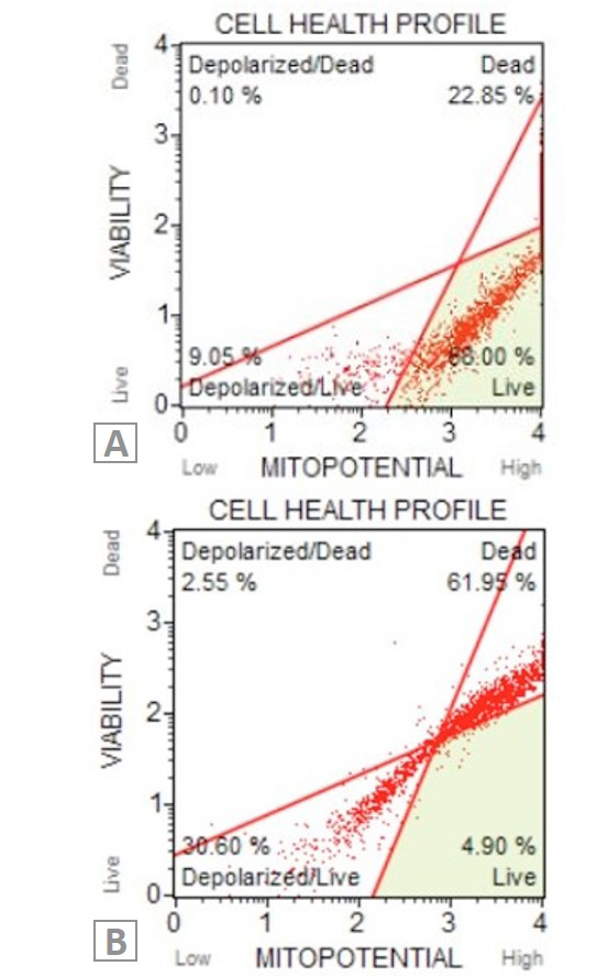
Analysing the mitochondrial membrane potential changes of A549 cells. A, Untreated A549 cells; B, A549 cells exposed to C2 ceramide.
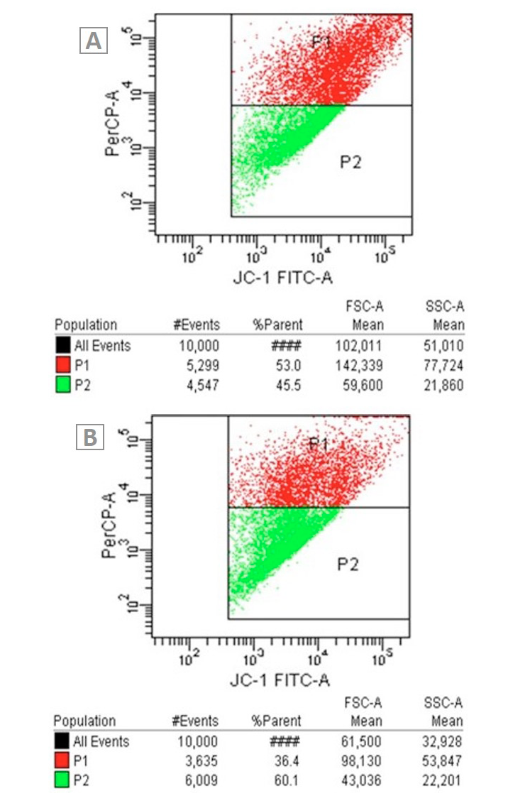
Mitochondrial membrane potential changes of A549 cells. A, Untreated A549 cells: B, A549 cells exposed to ceranib-2.
Ultrastructural changes of A549 cells. A, Untreated A549 cells: ∆, normal cell membrane; →, normal nuclear membrane. B, A549 cells exposed to C2 ceramide for 24 h: →, autophagic vacuols; ∆, loss of cristae. C, chromatin condensation and ◊, is circular shaped cell.
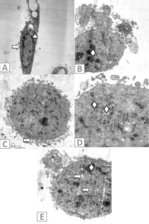
Ultrastructural changes of A549 cells. A, Untreated A549 cells: →, undamaged cell membrane; ∆, normal nuclear membrane. B, A549 cells exposed to ceranib-2 for 24 h. ◊, fragmented nucleus. C, →, Circular shaped cell and membrane blebbing. D, ◊, Lipid droplets in cells. E, ◊, DNA disintegration, →, holes in cell.


