Cocktail Phage Therapy for Bacteria Contaminating Meat in Egypt
Cocktail Phage Therapy for Bacteria Contaminating Meat in Egypt
Amany M. Reyad1*, Aya Maher Rabie1, Reda Mohamed Taha1 and Khalid El-Dougdoug2
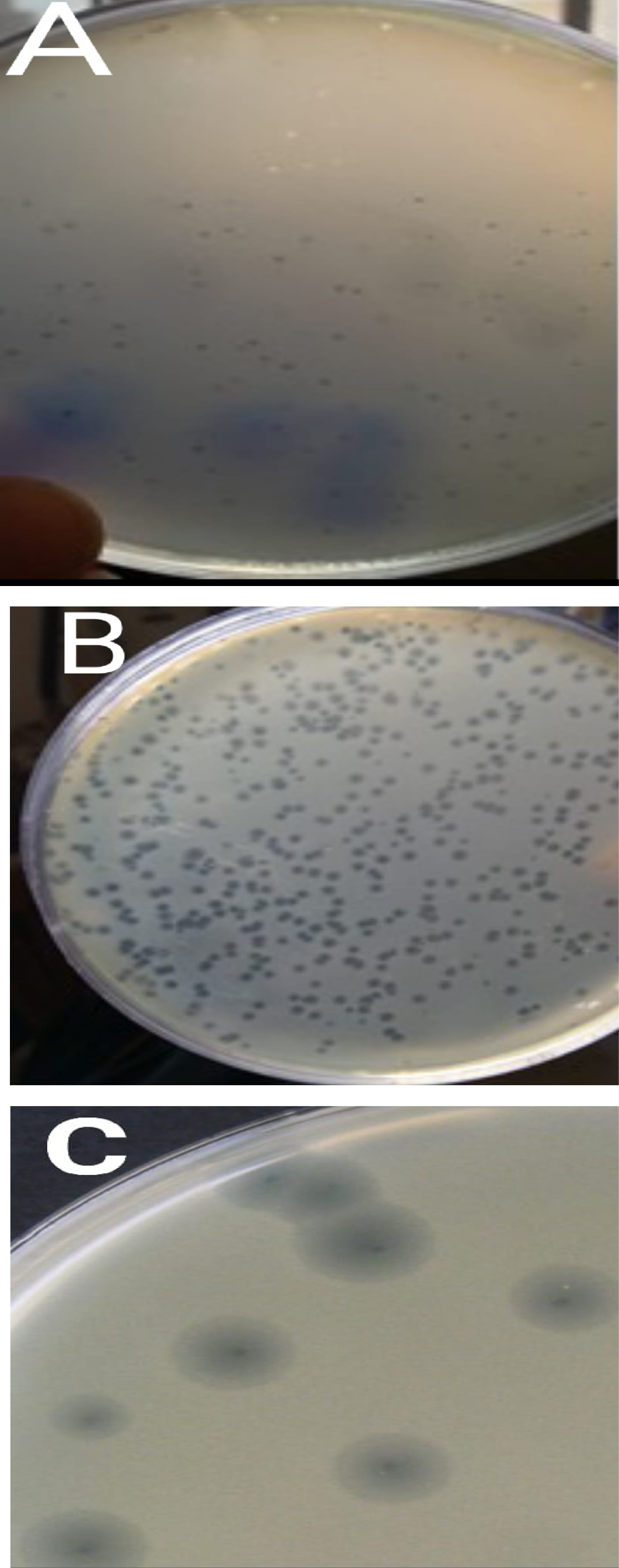
Plate photos showing A: S. aureus phage plaques, B: E. coli phages plaques and C: K. pneumoniae phage plaques. (A) The plaque is circular, clear, without center, without halo, and its size is 1.5mm. (B) E. coli phage 1: plaque zone Circular, clear, without center, without halo, and its diameter is 1mm. E. coli phage 2: Circular, turbid, without center, with Halo, and its diameter is 1.5 mm. (C) Circular, clear with center, with large halo, and its diameter is 6 mm.
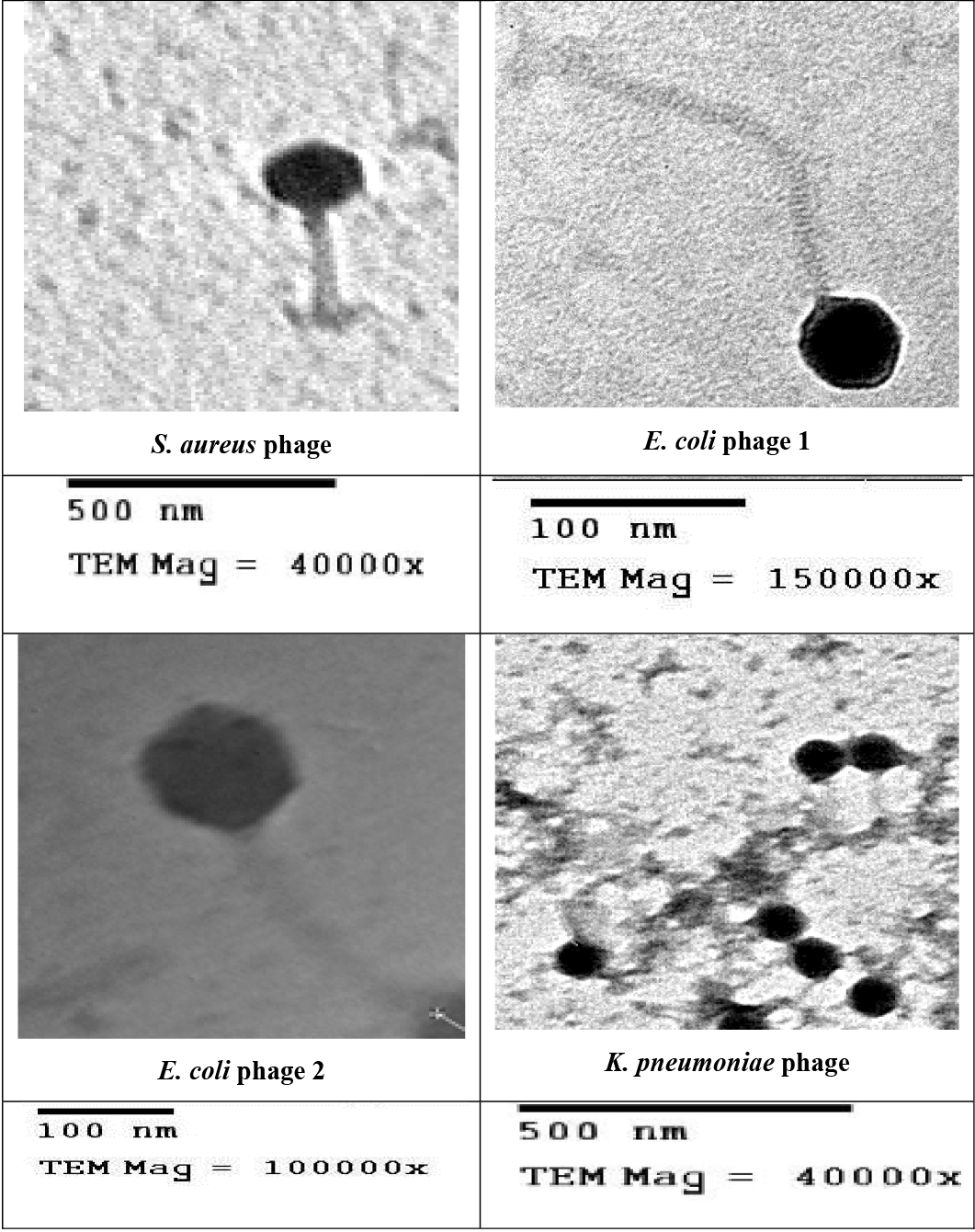
Electron micrographs of the four isolated phages. The magnification bar corresponds to 100 and 500 nm with high voltage equal to 80 KV.
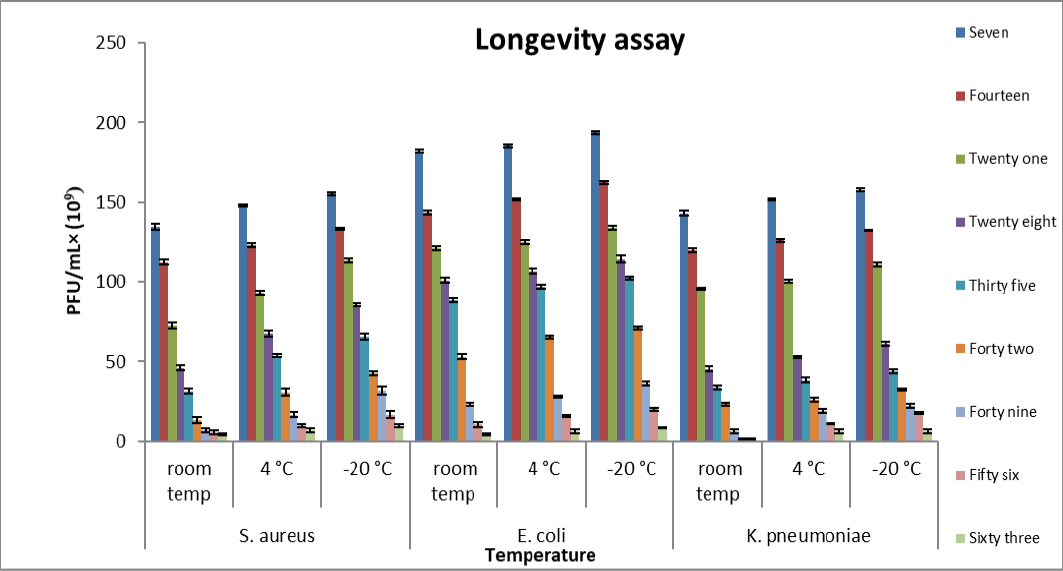
Histogram showing longevity of S. aureus, E. coli, and K. pneumoniae phages at different temperatures. Data are the means of the three replicates and error bars represent the standard errors of the means.


