
Cloning of the Gene Encoding Mycoplasma hyopneumoniae P36 Protein, Expression and Evaluating the Antigenicity of Recombinant Protein


Cloning of the Gene Encoding Mycoplasma hyopneumoniae P36 Protein, Expression and Evaluating the Antigenicity of Recombinant Protein
Nguyen Thi Thu Hien1, Dong Huu Rin2, Nguyen Xuan Hoa1, Le Viet Tuan Khanh2, Phung Thang Long1*, Dinh Thi Bich Lan1

PCR amplification of the gene encoding M. hyopneumoniae P36 protein from genomic DNA isolated from fresh lung tissue samples of PEP infected pigs in Thua Thien Hue province, Vietnam with specific P36 primer pairs. Lane M: DNA size marker (100-1500 bp, Bio Basic); Lanes 1-2: PCR products of the M. hyopneumoniae P36 gene.

Electrophoresis results of PCR products on gel agarose 1% to identify the presence of the gene encoding M. hyopneumoniae P36 protein and the recombinant pGEM®-T Easy/P36 vector in E. coli TOP 10. Lane M: DNA size marker (100-1500 bp, Bio Basic), Lanes 1, 3, 5: PCR products with specific primer pairs for M. hyopneumoniae P36 gene; Lanes 2, 4, 6: PCR products with M13 primer pairs of the pGEM®-T Easy vector.
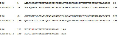
Comparison of amino acid sequence of recombinant P36 protein from M. hyopneumoniae with the amino acid sequence of recorded P36 protein of M. hyopneumoniae 7448 in GenBank (accession No. AAZ53511.1).

SDS-PAGE analysis of the expression of recombinant M. hyopneumoniae P36 protein in E. coli BL21(DE3) cells on LB medium. M: Protein weight marker (10-200 kDa, Bio Basic); Lane 1: Soluble P36 protein (in supernatant) of transformed E. coli BL21(DE3) cells induced with 0.8 mM IPTG when OD600 of the cultures reached 0.8 and incubated at 37oC, 150 rpm for 8 h post-induction; Lane 2: Inclusion body proteins of transformed E. coli BL21(DE3) cells in the same condition of expression; Lanes 3-4: Purified recombinant M. hyopneumoniae P36 protein.

Evaluation of antigenicity of recombinant M. hyopneumoniae P36 protein by ELISA. Wells 1-4 coated with the recombinant M. hyopneumoniae P36 protein (20µg/mL); NC: uncoated with the recombinant M. hyopneumoniae P36 protein (coating buffer).

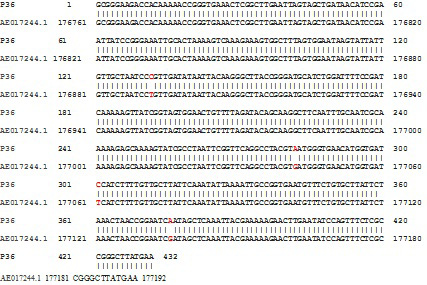
Comparison of nucleotide sequence of gene encoding P36 protein from M. hyopneumoniae isolated from PEP infected pigs in Thua Thien Hue province, Vietnam with the nucleotide sequence of recorded P36 gene of M. hyopneumoniae 7448 in GenBank (accession No. AE017244.1).

