
Clinical, Pathological and Molecular Investigations of Peste des Petits Ruminants Virus Infection in Goats from Turkana County in Kenya


Clinical, Pathological and Molecular Investigations of Peste des Petits Ruminants Virus Infection in Goats from Turkana County in Kenya
Simon Mwangi Kihu1*, George Chege Gitao1, Lily Caroline Bebora1, Njenga Munene John1, Gidraph Gachunga Wairire2, Ndichu Maingi1, Raphael Githaiga Wahome1, Davis Njuguna Karanja1, Julius Otieno Oyugi3, Ernest Lutomia3

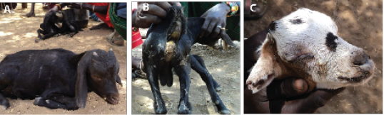
A. Goat suspected to have PPR infection in a depressed state, B. Diarrhoea and soiled anal region and hind legs, C. Showing muco-purulent ocular nasal discharges, matting of eye lids and encrusted.
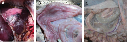
A. Inflamed lung showing hepatized apical lobe in goat, B. Hyperaeic intestinal mucosa showing hemorrhagic points, C. Swollen mesenteric lymph node (arrow) and congested mesenteric veins.
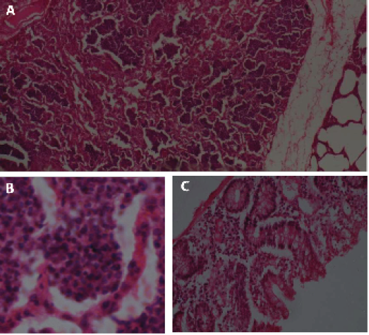
A. Lung of goat showing collapsed alveoli, B. infiltration with mononuclear cell in the alveoli and multinucleated syncitia, C. Large intestine showing proliferation of goblet cells and infiltration of inflammatory cells.


