
Clinical Evaluation and Pathological Findings of Air Rifle Shot in Slow Lorises (Nycticebus spp.) At The Animal Rehabilitation Center of Yayasan Inisiasi Alam Rehabilitasi Indonesia (YIARI) Bogor Regency


Clinical Evaluation and Pathological Findings of Air Rifle Shot in Slow Lorises (Nycticebus spp.) At The Animal Rehabilitation Center of Yayasan Inisiasi Alam Rehabilitasi Indonesia (YIARI) Bogor Regency
Hanita Fadhilla1, Shafia Khairani1,2*, Ahmad Fitrah3, Wendi Prameswari4, Nur Purba Priambada4, Indri Saptorini4, Imam Arifin4
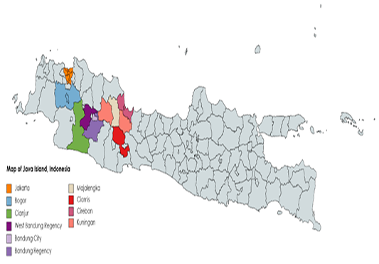
The area from which the slow lorises were rescued.

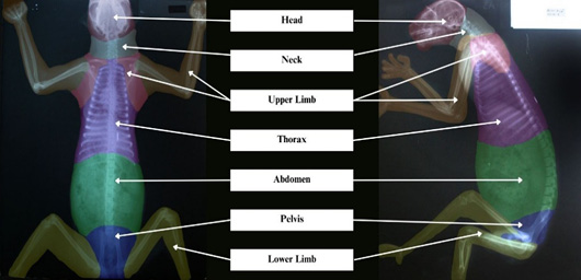
Anatomy region in slow loris radiography (YIARI, 2019).
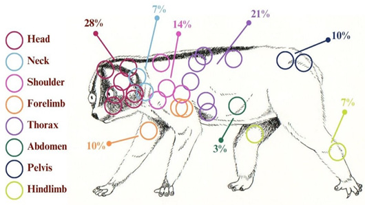
Schematic representation of pellet location findings.
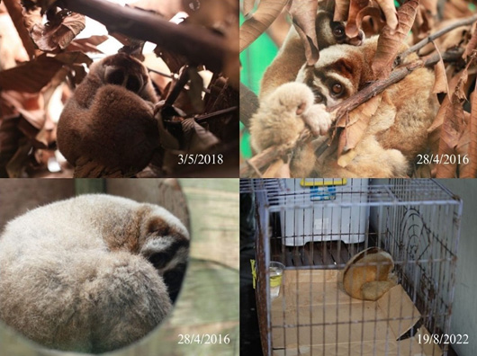
The rounded shape of the slow loris when sleeping (YIARI, 2016; 2018; 2022).
The slow lorises eyes are reflective due to the presence of the tapetum lucidum.

Intact pellet in the subcutaneous part of the head of individual 07J20 (A) and an intramuscularly intact pellet in the left forelimb of individual 11P15 (B) (YIARI, 2015; YIARI, 2022).
Encapsulated pellet in individual 04E22 (YIARI, 2022).

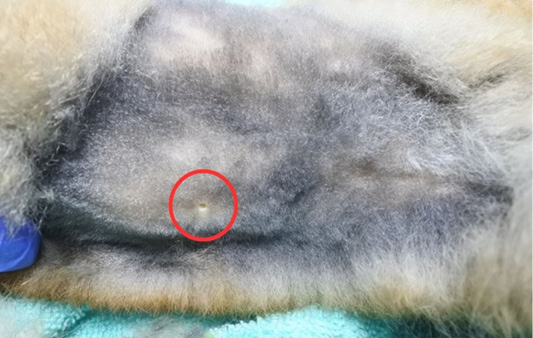
Entry wound in abdomen individual 02B20 (YIARI, 2020).
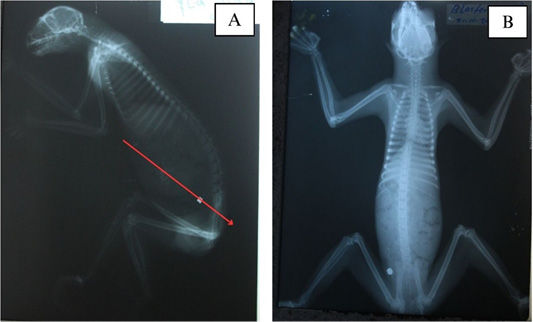
Radiographic results of lateral (A) and dorsoventral (B) views of individual 02B20. It can be seen that the pellet is pointing from the ventral to the dorsocaudal (YIARI, 2020).

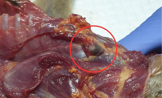
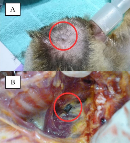
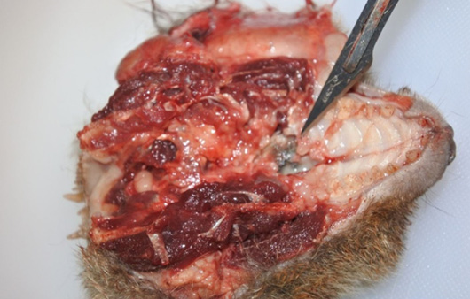
Necropsy documentation of head region individual 10M19 (YIARI, 2019).

Radiography image that was taken before (up) and after (down) surgery in individual 09L18.

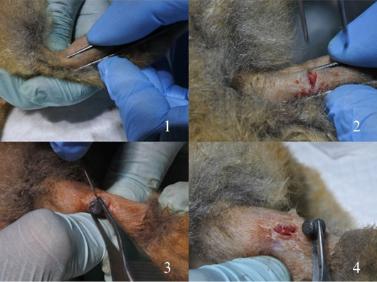
Surgical removal of a pellet lodged in the subcutaneous left lower extremity of individual 08K18 (YIARI, 2018).

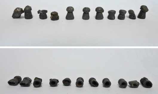
Some pellets were removed through surgery and stored in the medical record file (YIARI, 2018).


