

Biochemical and Histopathological Changes in Immune and Non Immune Broilers after Inoculation of Field Infectious Bursal Disease Virus


Biochemical and Histopathological Changes in Immune and Non Immune Broilers after Inoculation of Field Infectious Bursal Disease Virus
Beenish Zahid1,*, Asim Aslam2, Zafar Iqbal Chaudhry2 and Raheela Akhtar2
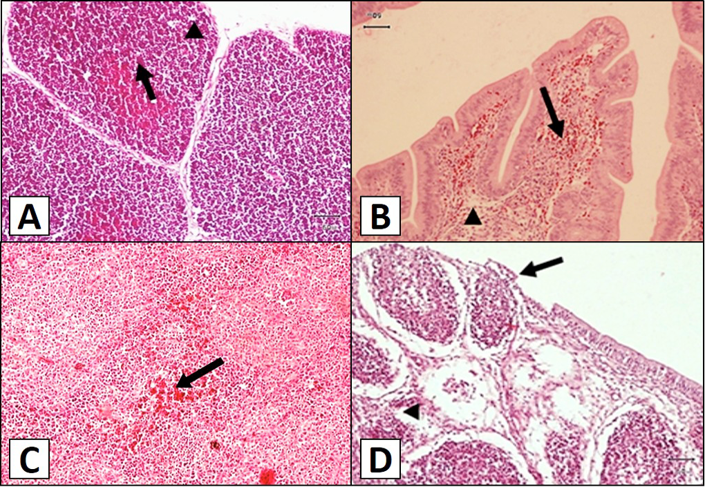
Fig 1
Histological structure of chicken thymus, bursa and spleen. A, thymus of group A at 3rd day post-infection (PI) showing hemorrhages (arrow) and mild lymphocytic depletion (arrowhead) in the follicles; B, Bursa of group A at 5th day PI showing severe hemorrhages (arrow) and lymphocytic depletion (arrowhead) in bursal follicles. C, spleen of group B at 5th day of PI showing severe congestion (arrow) and leukocytic infiltration (arrowhead); D, bursa from group A at 9th day of PI showing severe lymphoid follicle necrosis (arrowhead) with degraded epithelium (arrow).

August 2017
Vol. 49, Iss. 4, Pages 1151-1546

