Baohuoside I Inhibits Benzo(a)Pyrene-Induced Metastasis in A549 Lung Cancer Cells by Modulating STAT3 and EMT
Baohuoside I Inhibits Benzo(a)Pyrene-Induced Metastasis in A549 Lung Cancer Cells by Modulating STAT3 and EMT
Muhammad Khan1,*, Amara Maryam2, Hafiz Abdullah Shakir1, Javed Iqbal Qazi1 and Yongming Li3,*
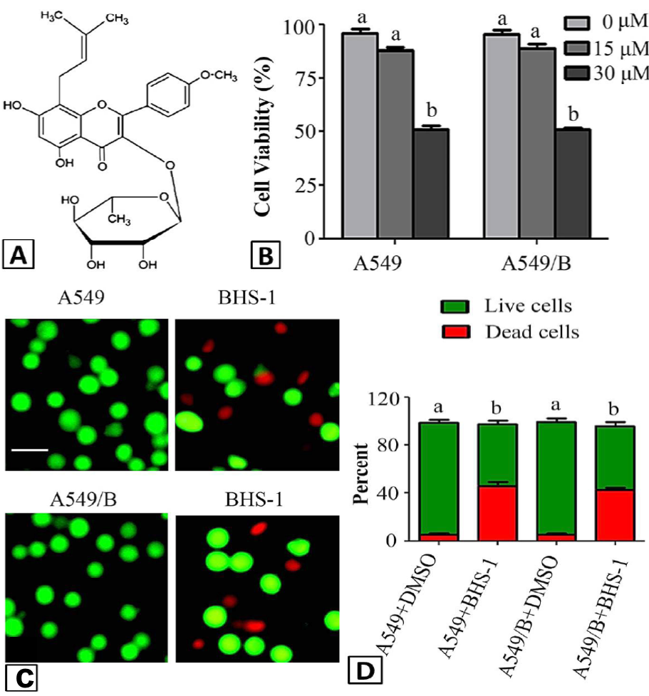
Effect of BHS-1 on proliferation and cell death of A549 and A549/B cells. A, Chemical structure of BHS-1; B, A549 and A549/B cells were treated with indicated concentrations of BHS-1 for 24 h and cell proliferation was determined by MTT assay. Data are expressed as Mean±SEM of 3 different experiments. Columns not sharing the same superscript letter differ significantly (P<0.05); C, A549 and A549/B cells were treated with 30 μM BHS-1 for 24 h and cell death was evaluated by live/dead assay; D, Data from live/dead assay are expressed as Mean±SEM of 3 different experiments. Columns not sharing the same superscript letter differ significantly (P<0.05).
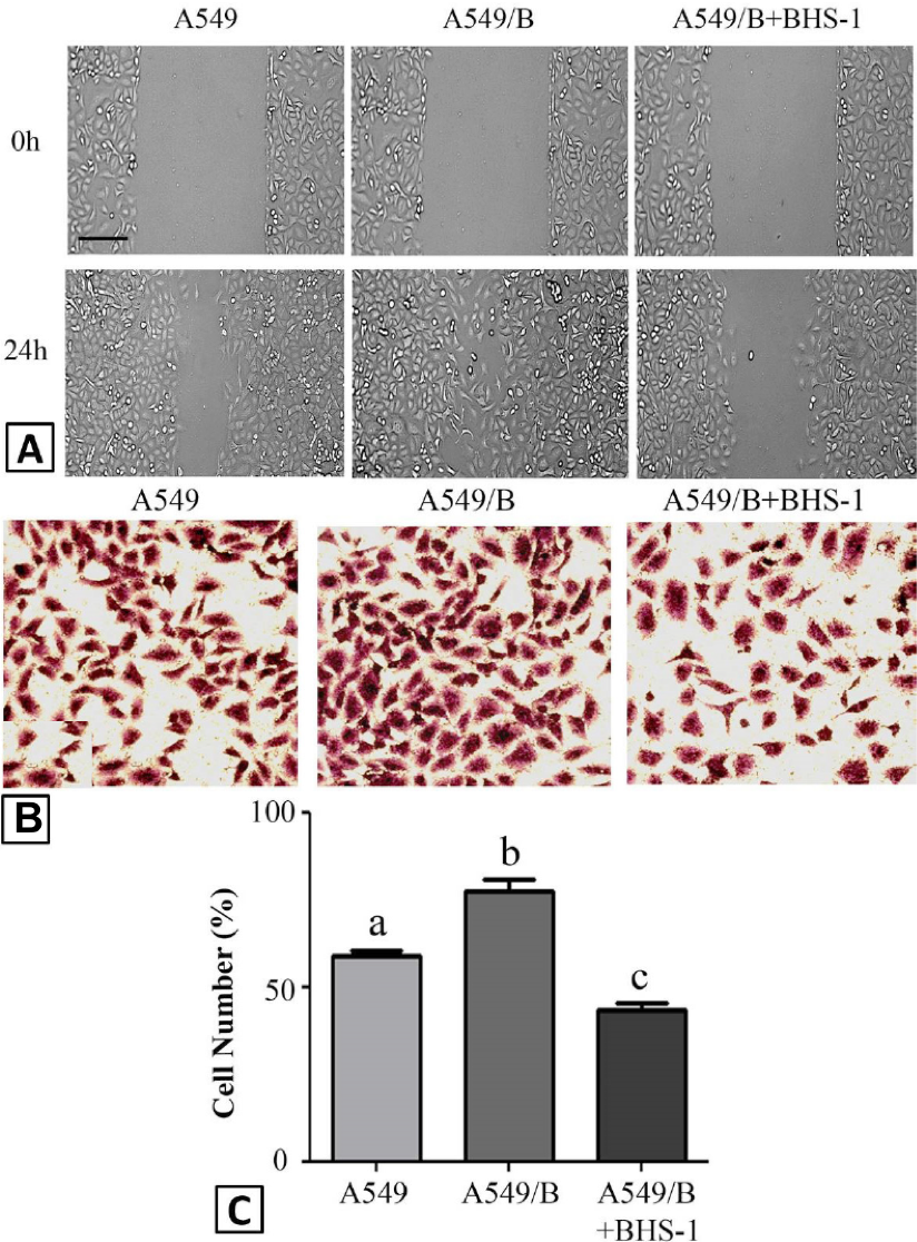
Effect of BHS-1 on BaP-induced cell migration: A, determination of migration ability of cells by wound healing assay as described in Materials and Methods section; B, determination of migration ability of cells by Transwell chamber assay as described in Materials and Methods section; C, statisctical analysis of data from Transwell chamber assay. Data from live/dead assay are expressed as Mean±SEM of 3 different experiments. Columns not sharing the same superscript letter differ significantly (P<0.05).
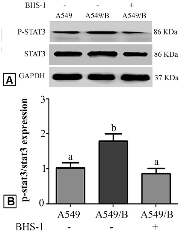
Western blot analysis of STAT3 activation: A, A549 and A549/B cells were treated with DMSO and BHS-1 (15μM) for 24 h and expression and phosphorylation of STAT3 was determined by Western blot analysis; B, protein expressions of p-STAT3/STAT3 from 3 different experiments. Columns not sharing the same superscript letter differ significantly (P<0.05).
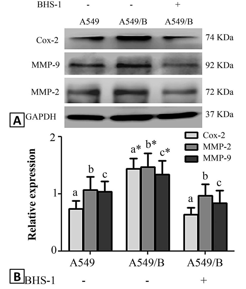
Modulatory effect of BHS-1 on expression of Cox-2, MMP-2 and -9 in response to chronic exposure of BaP: A, A549 and A549/B cells were treated with DMSO and BHS-1 (15μM) for 24 h and expressions of Cox-2, MMP-2 and -9 were determined by Western blot analysis; B, protein expressions from 3 different experiments were quantified by densitometry analysis. Similar superscript letters having * differ significantly with similar superscript letters without * at P<0.05.
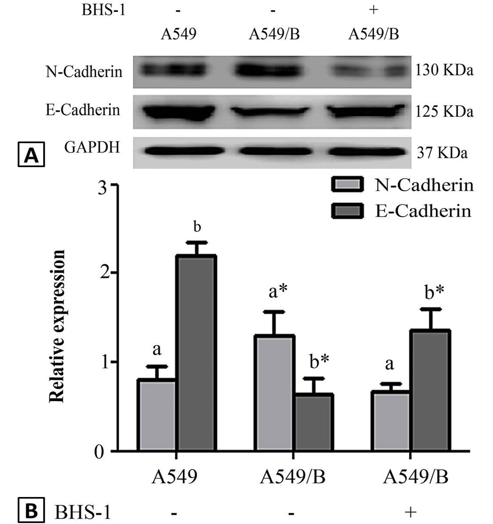
Modulatory effect of BHS-1 on BaP-induced EMT: A, A549 and A549/B cells were treated with DMSO and BHS-1 (15μM) for 24 h and expressions of N-cadherin and E-cadherin was determined by Western blot analysis; B, protein expressions from 3 different experiments were quantified by densitometry analysis. Similar superscript letters having * differ significantly with similar superscript letters without * at P<0.05.


