
Antischistosomal Activity of Trigonella foenum (Fenugreek) Crude Seeds Water Extract on Infected Mice


Antischistosomal Activity of Trigonella foenum (Fenugreek) Crude Seeds Water Extract on Infected Mice
Gamalat Y. Osman1, Elham M. Hassan1*, Tarek A. Salem2, Azza H. Mohamed1
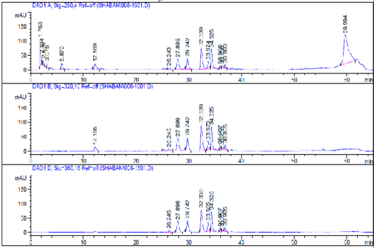
High performance liquid chromatography (HPLC) analysis of FWE analysis.

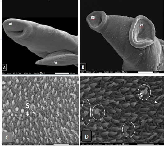
Scanning electron micrographs of S. mansoni worm collected from (A) Infected control mouse showing an anterior end with normal oral (os) and ventral suckers (vs). (B) Infected mouse treated with FWE showing no clear change in the oral and ventral suckers. (C) Enlarged portion of (A) showing oral sucker region covered with sharp spines. (D) Enlarged portion of (B) showing some corrosions in oral sucker spines after FEW treatments (circled).

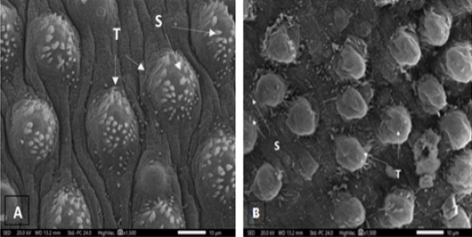
Scanning electron micrographs of S. mansoni worm collected from (A) Infected control mouse showing normal dorsal tegument tubercles (T) with spines (S). (B) Infected mouse treated with FWE showing mostly reduced spines (S) density and abnormal tubercular shape (T).
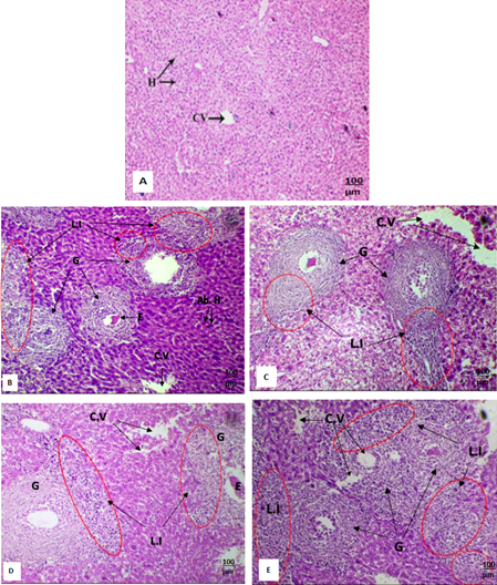
(A-E): Light micrograph of liver section of (A) Normal control mouse showing normal hepatocytes (H) and central vein. (B) S. mansoni infected mouse eight-week post infection showing abnormal hepatocytes (Ab. H), multiple Schistosoma eggs (E) provoke granuloma formation (G) of large diameters containing extended regions of lymphocytic infiltration (L.I). Cytoplasmic vacuolization is clear (C.V). (C) S. mansoni infected mice treated with PZQ drug showing granuloma (G) with high lymphocytic infiltration (L.I), clear cytoplasmic vacuolization (C.V). (D) S. mansoni infected mice treated with FWE showing granuloma (G) with clear lymphocytic infiltration (L.I) and mild cytoplasmic vacuolization (C.V). (E) S. mansoni infected mice treated with PZQ accompanied with FWE showing multiple granuloma (G) with high lymphocytic infiltration (L.I), multiple cytoplasmic vacuolization (C.V). (A-E: H and E X100).

