
Anatomical Variations of the Portal Vein in Ruminants


Anatomical Variations of the Portal Vein in Ruminants
Reda Mohamed1, 2*
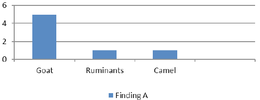
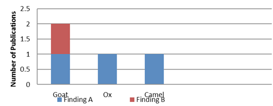
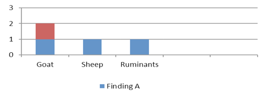
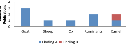
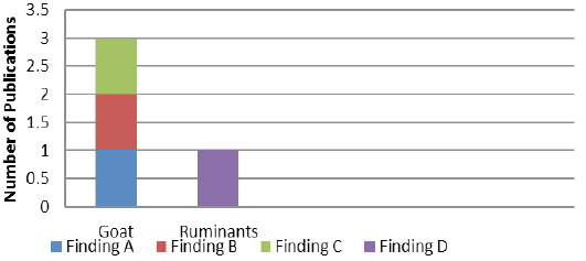
A graph showing the number of publications and the findings about the branches of the portal vein.

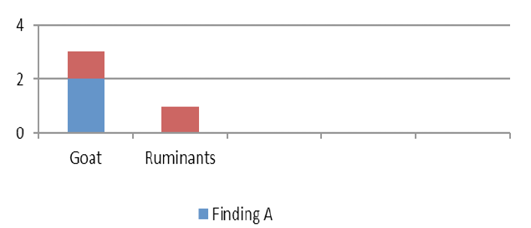
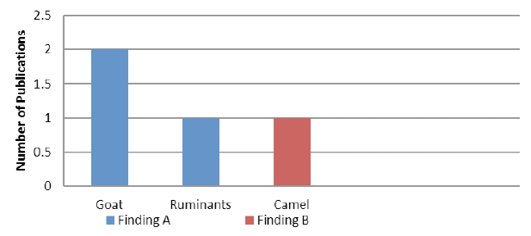
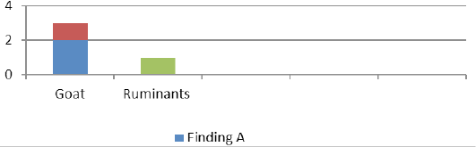
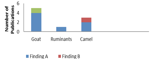
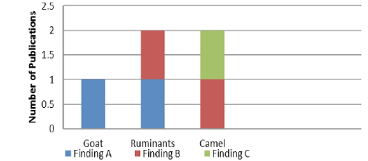
A graph showing the number of publications and the findings about the branches of the splenic vein.
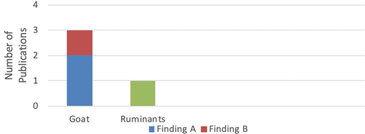
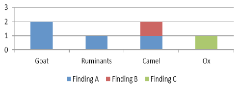
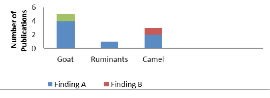
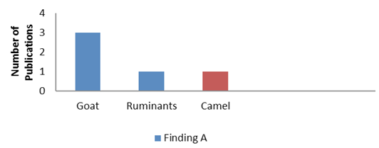
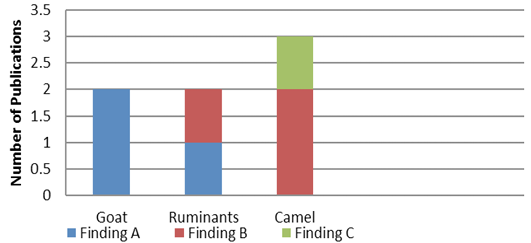
A graph showing the number of publications and the findings about the branches of the right ruminal vein.

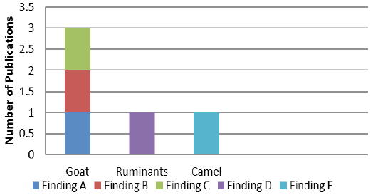
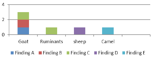
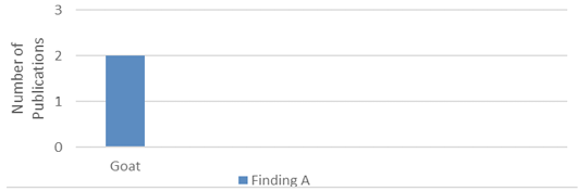
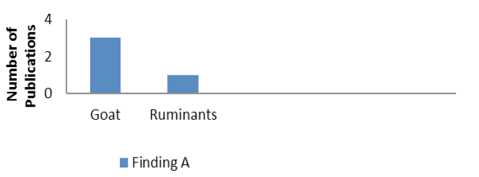
A graph showing the number of publications and the findings about the origin of the epiploic branch.

A graph showing the number of publications and the findings about the branches of the reticular vein.

A graph showing the number of publications and the findings about the branches of the left gastric vein.
A graph showing the number of publications and the findings about the branches of the left ruminal vein.

A graph showing the number of publications and the findings about the branches of the accessory reticular vein.

A graph showing the number of publications and the findings about branches of the left gastroepiploic vein.

A graph showing the number of publications and the findings about origin of the gastroduodenal vein.

A graph showing the number of publications and the findings about branches of the gastroduodenal vein.

A graph showing the number of publications and the findings about branches of the right gastric vein.

A graph showing the number of publications and the findings about branches of the right gastroepiploic vein.
A graph showing the number of publications and the findings about branches of the cranial mesenteric vein.

A graph showing the number of publications and the findings about the number of the jejunal veins.

A graph showing the number of publications and the findings about branches of the ileocolic vein.

A graph showing the number of publications and the findings about origin of the common trunk for colic rami and right colic veins.

A graph showing the number of publications and the findings about the origin of the caecal vein.
A graph showing the number of publications and the findings about branches of the caecal vein.
A graph showing the number of publications and the findings about branches of the caudal mesenteric vein.
A graph showing the number of publications and the findings about origin of the middle colic vein.


