

A Scanning Electron Microscopic Study of Argulus japonicus (Crustacea: Branchiura) Isolated from Goldfish (Carassius auratus) in Korea


A Scanning Electron Microscopic Study of Argulus japonicus (Crustacea: Branchiura) Isolated from Goldfish (Carassius auratus) in Korea
Mahanama De Zoysa1, Si-yun Ryu1, Hyeon-cheol Kim2 and Bae Keun Park1*
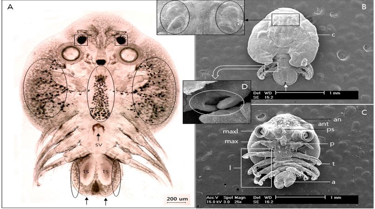
Fig 1
Whole body of male Argulus japonicus. A, Light micrograph. Note the melanophores patches (circles) scattered over dorsal surface of carapace lobes and abdomen. Compound eyes (square). Seminal vesicle (sv). Testis (ts). Note the rounded end of abdomen (arrows); B, Scanning electron micrograph of dorsal view. Compound eyes (circle in square). Abdomen separated by sinus (arrow); C, Scanning electron micrograph of ventral view. Not the pre-oral spine; D, Accessory copulatory projections on third leg (circle). a, abotomen; an, antennule; ant, antenna; c, carapace; l, swimming leg; max, maxilla; maxl, maxillule; p, proboscis; ps, preoral spine; sv, seminal vesicle; t, thorax; ts, testis.
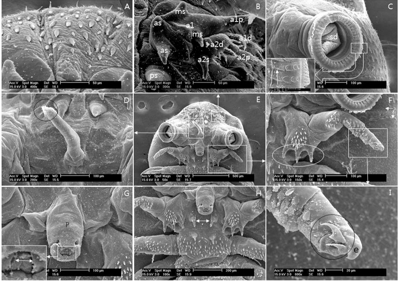
Fig 2
Scanning electron micrographs of anterio-ventral view of male A. japonicus. A, Dentate scales that face towards posterior that occur along the edge of the ventral carapace; B, Antennule and antennae; C, First maxilla. Rim of sucker with supporting rods and sclerites. Basis elongated plate (arrow in square); D, Pre-oral spine, Note the terminal (circle); E, Anterio- ventral part; F, Maxilla with five segments, basal podomere (plate) with three tooth (circle). Two lobes of respiratory area (arrows); G, Proboscis without any dorsal scales. Mouse tube (arrow in square); H, Basement of maxillae and first leg. Basal plate scales of maxillae (arrow); I, Terminal segment of maxilla in a blunt fused extension has two recurved sharp claws (Circle). e, extension; a, antenna; as, anterior spine; a1, first antenna; a1d, first antenna distal part; a2d, second antenna distal part; a2p, second antenna proximal part; a2s, spine of second antenna; ms, median spine; p, proboscis; ps posterior spine; a1p, first antenna proximal hook; sd, suckiong disc.

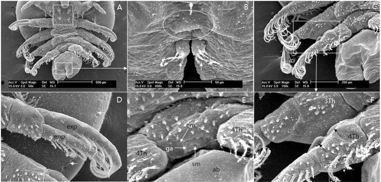
Fig 3
Scanning electron micrographs of postero-ventral view of male A. japonicus. A, Thorax and abdomen. Spines of each post maxillary spine (thoracic spine?) (square); B, Anus (circle). Caudal rami (arrow). Note the four setae; C, Third and fourth leg; D, First swimming leg; Exopodium and endopodium with plumose setae. Posterior region of respiratory area (arrows); E, Male’s genital aperture to show spermatophore secretion; F, Peg (arrow) on the right fourth leg and the socket of right third leg. Note the socket (indentation) on the ventral side of forth leg. ab, abdomen; cm, contracted muscle; exp, exopodium; enp, endopodium; ga, genital aperture; sm, spermatophore; 3Th, third leg; 4Th, fourth leg.

April 2017
Vol. 49, Iss. 2, Pages 425-759

