
During year 2015, a survey was conducted in 18 golf course greens from nine states (Selangor, Negeri Sembilan, Melaka, Johor, Pahang, Perak, Kedah, Pulau Pinang and Wilayah Persekutuan) of Peninsular Malaysia for nematode, Meloidogyne graminis infections on the turf grasses. Samples were collected based on the sparsely growth and chlorotic appearance of the greens. A total of 36 soils and roots sample were collected. Scanning electron microscopy (SEM) was used to identify the parasitic nematode. Both the field symptoms and SEM micrographs confirmed that the nematode isolate was M. graminis. Since this nematode has been known to damage the greens and other plants in other part of the world then the probability for the specie to adapt to other hosts other than the family of poaceae under Malaysian climate should not be discounted. Therefore, screening and restriction of movement of planting materials be observed critically.
Short Communication · Volume 51, Issue 4, 5 June 2019 · Open Access
Ultrastructural Characterization of Meloidogyne graminis from Golf Course Turf Grasses in Peninsular Malaysia
· · · · ·
Affiliations & Notes ⌄
- 1Department of Plant Protection, Faculty of Agriculture, Universiti Putra Malaysia 43400, Serdang, Selangor, Malaysia 2Center for Insect Systematic, School of Environmental and Natural Resource Sciences, Faculty of Science and Technology, Universiti Kebangsaan Malaysia 43600 Bangi, Selangor, Malaysia 3Department of Plant Pathology, University College of Agriculture, University of Sargodha, Sargodha 40100, Punjab 4Plant Pathology Research Institute, Ayub Agricultural Research Institute, Faisalabad
Article Info ⌄
- Journal:
- Pakistan Journal of Zoology
- Published:
- 5 June 2019

From the article
- Saccate M. graminis females were observed partially embedded in the root tissue (Fig. 1A).
- For most of the oval shaped females, lateral posterior half of the body is seen outside the root while the remaining half of the body is embedded in the cortical tissue of the root.
- However, the cortical tissue around the body of the nematode seems to split longitudinally to accommodate the swollen mature female (Fig. 1B).
- Contrary, the spherical shaped females have their head and neck protruded to only one side of the body, at an angle with longitudinal axis of the body (Fig. 2).
Selected from the article's own text.
Abstract
Materials and methods
In 2015, survey was conducted in 18 golf course greens from 9 states (Selangor, Negeri Sembilan, Melaka, Johor, Pahang, Perak, Kedah, Pulau pinang and Wilayah Persekutuan) of Peninsular Malaysia for M. graminis infections on the turf grasses. Samples were collected based on the sparsely growth and chlorotic appearance of the greens. A total of 36 soils and root samples were collected following the sampling procedure proposed by Speijer and De Waele (1997). Roots were separated from soil, washed and stained by boiling in acid fuschin lactophenol, following Hooper (1986). Root galls with matured females were cut into 1cm length, fixed in formalin acetic acid, post fixed in 1% buffer osmium tetraoxide and then dehydrated through sequence of graded ethanol to absolute ethanol. The samples were then dried in critical point drier (CPD) Balzar 030, mounted on stub and splutter gold coated and viewed under JOEL 6400 at accelerated voltage of 15 kv.
Results
Saccate M. graminis females were observed partially embedded in the root tissue (Fig. 1A). Manifest symptoms of infested roots were slight enlargement of the root where the female nematode resides. The extent of body exposure outside the root tissues is influenced by the shape of the swollen mature female. For most of the oval shaped females, lateral posterior half of the body is seen outside the root while the remaining half of the body is embedded in the cortical tissue of the root. However, the cortical tissue around the body of the nematode seems to split longitudinally to accommodate the swollen mature female (Fig. 1B). Only the head and neck of the spherical females were found embedded in the root tissue, the enlarged part of the body remaining outside the root in egg laying females, the exposed posterior part of the body was generally covered by eggs and gelatinous matrix (Fig. 1B).
The oval-shaped females have their vulva and anus located on protuberance on the same longitudinal axis of the body at opposite end of the head (Fig. 1C). Contrary, the spherical shaped females have their head and neck protruded to only one side of the body, at an angle with longitudinal axis of the body (Fig. 2).
The perineal pattern (posterior cuticular pattern) of the nematode was found to be oval shaped, possessing trapezoidal dorsal arch. The striae were smooth or slightly wavy with prominent lateral lines (Fig. 3).
Discussion
Of the 18 golf courses sampled, M. graminis was found in 14 golf courses across peninsular Malaysia. Our results reveal how widely distributed the parasite is on golf course greens in Peninsular Malaysia. Our findings corroborate with studies from other workers ; ; who reported M. graminis as an economically important species to golf course industries through decline in turf quality by causing the grass to be chlorotic, stunted in growth and at some levels may cause the plant to die. Similarly, Hunt and Handoo (2009) found that the most economically damaging nematode species on horticultural
and field crops are the root knot nematodes. Having a worldwide distribution, and being obligate, make them parasites of the roots of many plant species, of monocots and dicots, and woody and herbaceous plants. Obvious sign of RKN infection are root galls, shoot chlorosis, stunted growth, nutrient deficiencies, and paving way for secondary infections by other pathogens . A high level of damage can lead to total crop loss. So far, there are nine species of Meloidogyne associated with turf grasses worldwide, including M. graminis Sledge and Golden Whitehead, M. chitwoodi Golden, M. incognita Kofoid and White, 1919, M. fallax, M. Graminicola Golden and Birchfield, M. marylandi Jepson and Golden, M. Microtyla Mulvey, M. minor Karssenand M. naasi Franklin ; ; . This is the first time this nematode is reported in Malaysia. There is every possibility that the nematode could have been transported with the stolon planting materials from United States. However, the probability for the specie to adapt to other hosts other than the family of poaceae under Malaysian climate should not be discounted. Therefore, screening and restriction of movement of planting materials be observed critically.
Statement of conflict of interest
The authors declare no conflict of interest.
Figures
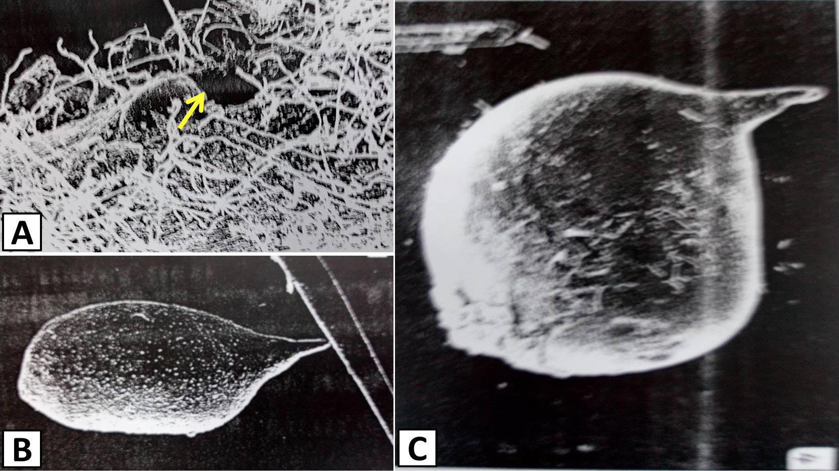
Turf grass root infected by female (A) and an oval-shaped saccate female (B) of Meloidogyne graminis. C, oval shaped female detached from root tissues. The head is on the opposite side of perineal pattern along the body axis.
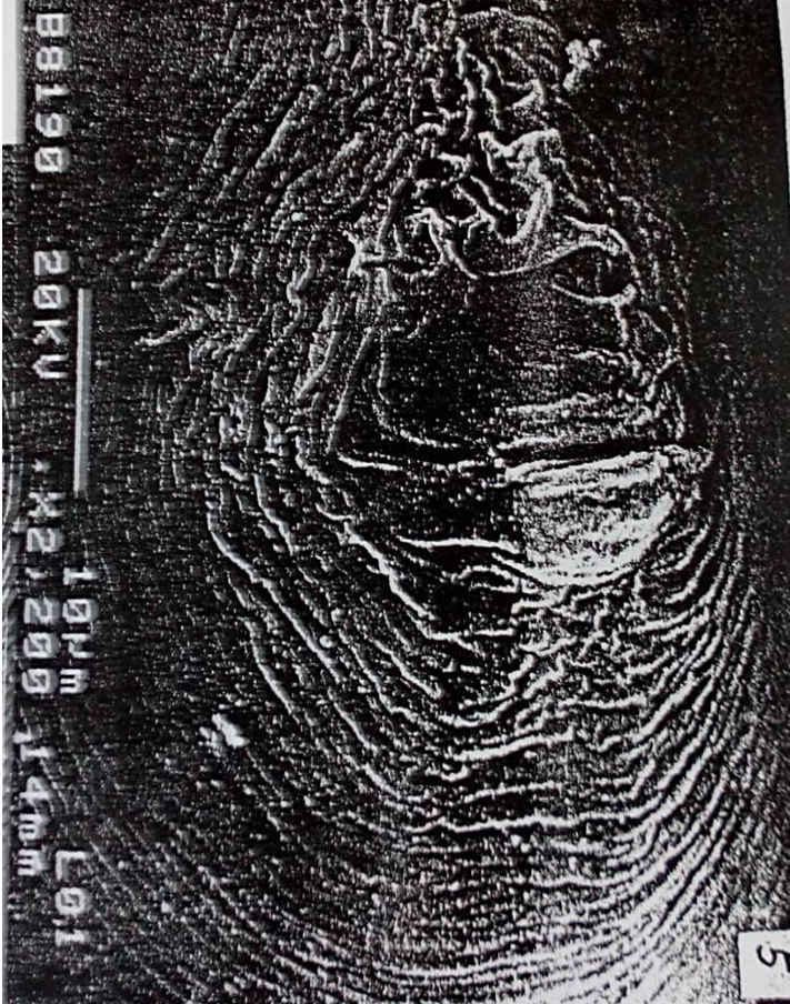
Spherical-shaped body of the saccate female of Meloidogyne graminis. The neck region is almost at right angle to the longitudinal body axis.
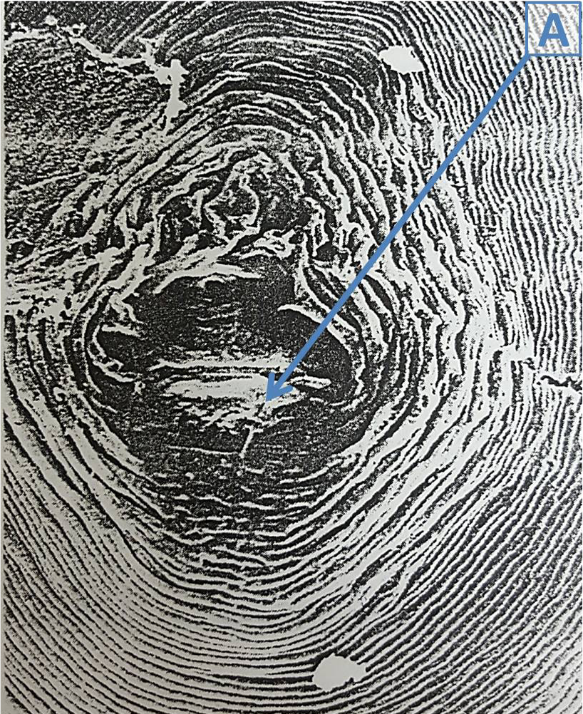
A side view of the vulva and anus on protuberance at the posterior terminal of the body.
References
- Bekal, S. and Becker, O.J., 2000a. Pl. Dis., 84: 1081-1084. https://doi.org/10.1094/PDIS.2000.84.10.1081[CrossRef]
- Bekal, S. and Becker, O.J., 2000b. Hortscience, 35: 1276-1278.
- Blake, J.H. and Doubrava, N., 1999. Root-Knot nematodes in the vegetable garden. Clemson Extension HGIC 2216. Online available at: http://hgic.clemson.edu/factsheets/HGIC2216.html
- Bongers, T. and Bongers, M., 1998. Appl. Soil Ecol., 10: 239-251. https://doi.org/10.1016/S0929-1393(98)00123-1[CrossRef]
- Cassida, K.A., Kirkpatrick, T.L., Robbins, R.T., Muir, J.P., Venuto, B.C. and Hussey, M.A., 2005. Nematropica, 35: 1-10.
- Coates-Beckford, P.L. and Malek, R.B., 1982. Nematropica, 12: 7-14.
- Cheng, Z., Grewal, P.S., Stinner, B.R., Hurto, K.A. and Hamza, H.B., 2008. Appl. Soil Ecol., 38: 174-184. https://doi.org/10.1016/j.apsoil.2007.10.007[CrossRef]
- Crow, W.T. and Grewal, P.S., 2009. Appl. Soil Ecol., 42: 107-117. https://doi.org/10.1016/j.apsoil.2009.02.005[CrossRef]
- Crow, W.T., 2005. Outlooks Pest Manage., 16: 10-15. https://doi.org/10.1564/16feb04[CrossRef]
- Crow, W.T. and Welch, J.K., 2004. Nematropica, 34: 31-37.
- Crow, W.T., Cuda, J.P. and Stevens, B.R., 2009. J. Nematol., 41: 217-220.
- Dunn, J. and Diesburg, K., 2004. Turf management in the transition zone. John Wiley & Sons, Inc., Hoboken, New Jersey.
- Franklin, M.T., 1965. Nematologica, 11: 79-86. https://doi.org/10.1163/187529265X00500[CrossRef]
- Giblin-Davis, R.M., Busey, P. and Center, B.J., 1995. J. Nematol., 27: 472-477.
- Griffin, G.D., Inserra, R.N. and Vovlas, N., 1984. J. Nematol., 16: 399-402.
- Grisham, M.P., Dale, J.L. and Riggs, R.D., 1974. Pythopathology, 64: 1485-1489. https://doi.org/10.1094/Phyto-64-1485[CrossRef]
- Hooper, D.J., 1986. Preserving and staining nematodes in Plant tissue. In: Laboratory methods for work with plant and soil nematodes, 6th edition (eds. J.F. Southey). HMSO, London, pp. 81-85
- Hunt, D. and Handoo, Z.A., 2009. Taxonomy, identification and principal species. In: Root-knot nematodes (eds. R.N. Perry, M. Moens and J.L. Starr). CABI, Publishing, UK, pp. 55-97.
- Khan, M.W. (ed.), 1993. Nematode interactions. Chapman & Hall, London, pp. 377. https://doi.org/10.1007/978-94-011-1488-2[CrossRef]
- McClure, M.A, Nischwitz, C., Skantar, A.M., Schmitt, M. and Subbotin, S.A., 2012. Pl. Dis., 96: 635-647. https://doi.org/10.1094/PDIS-09-11-0808[CrossRef]
- Mai, W.F. and Lyon, H.H., 1975. Pictoral key to genera of plant parasitic nematodes. Cornell University Press, Ithaca, NY, pp. 219.
- Milesi, C., Running, S., Elvidge, C., Dietz, J., Tuttle, B. and Nemani, R., 2005. Environ. Manage., 36: 426-438. https://doi.org/10.1007/s00267-004-0316-2[CrossRef]
- Mulvey, R.H., Townshend, J.L. and Potter, J.W., 1975. Canadian J. Zool., 53: 1528-1536. https://doi.org/10.1139/z75-188[CrossRef]
- Nischwitz, C., Skantar, A., Handoo, Z.A., Hult, M.N., Schmitt, M. and Mcclure, M.A., 2013. Pl. Dis., 97: 1424-1430. https://doi.org/10.1094/PDIS-03-13-0263-RE[CrossRef]
- Nyczepir, A.P., 2011. Nematropica, 41: 45-51.
- Rahman, M.L. and Evans, A.A.F., 1988. Nematology, 33: 451-459. https://doi.org/10.1163/187529287X00100[CrossRef]
- Subbotin, S.A., Inserra, R.N., Marais, M., Mullin, P., Powers, T.O., Roberts, P.A., Berg, E.V.D., Yeates, G.W. and Baldwin, J.G., 2011. Nematology, 13: 333-345. https://doi.org/10.1163/138855410X520936[CrossRef]
- Schwartz, B.M., Kenworthy, K.E., Crow, W.T., Ferrell, J.A., Miller, G.L. and Quesenberry, K.H., 2010. Crop Sci., 50: 723-729. https://doi.org/10.2135/cropsci2008.11.0672[CrossRef]
- Sikora, E.J., Guertal, E.A. and Bowen, K.L., 1999. Highlights Agric. Res., 46: 10-11.
- Sledge, E.B. and Golden, A.M., 1964. Proc. helminthol. Soc. Wash., 31: 83-88.
- Speijer, P.R. and de Waele, D., 1997. Screening of Musa germplasm for resistance and tolerance to nematodes. INIBAP Technical Guidelines 1. INIBAP, Montpellier, France, pp. 42.
- Starr, J.L., Ong, K.L., Huddleston, M. and Handoo, Z.A., 2007. Nematropica, 37: 43-49.
- Vandenbossche, B., Viaene, N., Sutter, N., de Maes, M., Karssen, G. and Bert, W., 2011. Nematology, 13: 245-256. https://doi.org/10.1163/138855410X517084[CrossRef]
- Wetzel, H.C., 2000. Nematode damage and management in lawns. Turfgrass Disease Information. Note 2 (TGIN-002). College of Agriculture and Life Sciences. Plant Pathology Extension. North Carolina State University. Available at: http://www.turffiles.ncsu.edu/Keywords/management.aspx#AR004342
- Ye, W., Zeng, Y., Tredway, L., Martin, S., Martin, M. and Fouly, H., 2012. Plant-parasitic nematodes in Carolina Turfgrass. Publication of the Carolinas Golf Course Superintendents Association, Carolinas Green, pp. 24-26.
Services
Article usage statistics
Loading…