
The purpose of this study was to investigate the effect of 5-fluorouracil (5-FU) on the expression of TGF-β1, cyclinD1, and CDK4 in fibroblasts of miniature pig bile duct scar model cultured in vitro. The bile duct scar fibroblasts were replicated by incision and anastomosis in miniature pigs and bile duct scar fibroblasts were obtained. After cell identification, the bile duct scar fibroblasts were given different concentrations of 5-FU (0, 0.001, 0.01, and 0.1 mmol/L). After 48 h of intervention, CCK-8 method was used to detect the cell proliferation level of each group; Western blot was used to detect the expression of TGF-β1, cyclinD1, and CDK4 protein in each group of cells; and real-time polymerase chain reaction was used to detect the expression levels of TGF-β1, cyclinD1, and CDK4 mRNA in each group of cells. The results showed that after 5-FU intervention, the proliferation of bile duct scar fibroblasts was inhibited, and the expressions of TGF-β1, cyclinD1, CDK4 mRNA and protein in the cells were down-regulated (P<0.05), showing concentration-dependence. In conclusion, 5-FU may have a role in inhibiting bile duct scars. One of the mechanisms may be related to its ability to inhibit the expression of TGF-β1, cyclinD1, and CDK4 in bile duct scar fibroblasts.
Short Communication · Volume 53, Issue 2, 27 February 2021 · Open Access
The Inhibition of Cell Proliferation of Bile Duct Scar Fibroblasts in Miniature Pigs by 5-fluorouracil and its Mechanism
· · · · · · ·
Affiliations & Notes ⌄
- 1Medical Faculty, Affiliated Hospital of Kunming University of Science and Technology, Kunming, Yunnan, 650504, China 2Department of Hepatobiliary and Pancreatic Surgery, Affiliated Hospital of Kunming University of Science and Technology The First People’s Hospital of Yunnan Province, 650032, China , * Corresponding author: virwm3@163.com
Article Info ⌄
- Journal:
- Pakistan Journal of Zoology
- Published:
- 27 February 2021

Abstract
Materials and methods
Four healthy male experimental miniature pigs of clean grade were selected and randomized into a normal control group and a bile duct scar model group, with 2 pigs in each group. The pigs were put in fast for 12 h before the operation. They were intravenously anesthetized with 2.5% sodium pentobarbital solution (45 mg/kg body weight). A midline incision was made in the upper abdomen, the skin and subcutaneous tissue were cut, and the common bile duct was exposed. In the bile duct scar model group, the common bile duct with a length of about 1 cm was freed near the duodenum. It was cut transversely for about 1/3 of its circumference, and was sutured with Dexon thread. The abdomen was sutured layer by layer. In the normal control, the abdomen was sutured after opening, without opening the common bile duct. Both groups were fed as routines after 12-h fasting. The bile duct tissues of the bile duct scar model group and the normal control group were taken one week after the operation, washed with phosphate buffer solution (PBS) three times, and cut into 2 mm3 tissue pieces, transferred to the culture flask and placed in a 5% carbon dioxide incubator at 37°C. After 4-h incubation, the cells adhered well, and DMEM medium (containing 20% inactivated fetal bovine serum, 100 U/mL penicillin, and 100 μg/mL streptomycin sulfate) was added to subculture the adherent cells. Fibroblasts were isolated and purified using differential adherence, and passaged when the cells grew to 90% confluence. Vimentin cells were immunofluorescence-positive, and cytokeratin cells were immunofluorescence-negative. The third-generation fibroblasts that fit the characteristics of fibroblasts were used for this experiment.
The third-generation fibroblasts of the normal bile duct from the normal control miniature pigs were selected as Group A. The third-generation fibroblasts of miniature pig bile duct scar in the bile duct model group were selected as Group B (0 mmol/L 5-FU), Group C (0.001 mmol/L 5-FU), Group D (0.01 mmol/L 5-FU) and Group E (0.1 mmol/L 5-FU).
CCK-8 method to detect cell proliferation.Keloid fibroblasts (cell density 2×10⁴/well) in the logarithmic growth phase of Groups A, B, C, D and E were inoculated into 96-well culture plates (100 μL/well). After incubating for 24 h in a 5% carbon dioxide incubator at 37°C, the 5-FU with the corresponding concentration was added to incubate for 48 h according to the different groups (the same amount of sterile physiological saline was added to Group A), and the CCK-8 reagent solution was added to incubate at 10 μL/well for 2.5 h. The culturing was then terminated. It was shaken up. The optical density (OD) value of each well was measured at 450 nm wavelength on an automatic quantitative microplate reader. The background deduction strategy was used to eliminate errors. Each experiment was repeated three times, and the average OD value was calculated for comparison. A higher OD value indicated stronger proliferative activity of the cell.
Expression levels of TGF-β1, cyclinD1, and CDK4 protein were detected by Western blot method. The cells in the logarithmic growth phase of each group were used for the experiment, and the total RNA of each group of cells was extracted. Proteins were separated by SDS-PAGE and transferred to PVDF membranes. After blocking overnight with 5% skim milk TBST solution, the membranes were washed, and the corresponding primary antibodies TGF-β1, cyclinD1, and CDK4 (1:500) were added, and the secondary antibodies Rabbit Anti-Mouse IgG secondary antibody (HRP) (1:5000) were added. They were developed and analyzed on a chemiluminescence imager after color development by ECL, with β-actin as an internal reference. The experiment was repeated three times to ensure the accuracy of the experimental results.
Semi-quantitative RT-PCR was used to analyze the expression of TGF-β1, cyclinD1, and CDK4 mRNA in each group of cells. The amplification conditions were predenaturation at 94°C for 5 min, denaturation at 94°C for 30 s, extension at 72°C for 1 min, and cycling 30 times, and finally extension at 72°C for 8 min to terminate the reaction. The forward primer of TGF-β1 is 5’-GCCCTGG-ACACCAACTATTGC-3’, and the reverse primer is 5’-GCACTTGCAGGAGCGCA-3’. The forward primer of cyclinD1 is 5’-GAGACCATCCCCCTGACGGC-3’, and the reverse primer is 5’-TCTTCCTCCTCCTCGGC-GGC-3’.The forward primer of CDK4 is 5’-TGATGCGCCAGTTTCTAAGAGG-3’, and the reverse primer is 5’-GGTCGGCTTCAGAGTTTCCACA-3’.The forward primer of β-actin is 5’-GACAACGGCTCC-GGCATGTGCA-3’, and the reverse primer is 5’-TGAG-GATGCCTCTCTTGCTCTG-3’. The ABI Stepone software v 2.3 qRTPCR system was used to collect data and analyze the results. The melting curve was a single peak type, indicating good primer specificity. With β-actin as an internal reference, the relative expression of the target gene was calculated using the 2-∆∆Ct method. The experiment was repeated three times and the average was taken.
Data analysis was performed using SPSS 25.0 statistical software (SPSS Inc., Chicago, IL, USA). The measurement data were expressed as mean ± standard deviation. Comparisons among multiple groups were analyzed by one-way variance with LSD-t test. The P<0.05 indicated that the difference was statistically significant.
Results and discussion
After 5-FU concentration gradient acted on bile duct scar fibroblasts, the OD value decreased significantly as the 5-FU concentration increased, indicating a significant increase in inhibition, and the difference was statistically significant (P<0.05; Fig. 1). It is suggested that 5 -FU can inhibit the proliferation of bile duct scar fibroblasts in miniature pigs. This conclusion is basically consistent with the results reported in previous study .
Compared with Group A, the expression levels of TGF-β1, cyclinD1, CDK4 protein and mRNA in Groups B, C, D and E were increased (P <0.05; Figs. 2, 3). It is suggested that the production of TGF-β1, cyclinD1 and CDK4 may play an important role in the process of bile duct scar formation in miniature pigs.
With the increase of 5-FU concentration, the expression levels of TGF-β1, cyclinD1, CDK4 protein and mRNA in cells decreased significantly (P<0.05; Figs. 2, 3), showing concentration-dependence. It is speculated that the inhibition of the proliferation of bile duct scar fibroblasts in miniature pigs by 5-FU is related to the regulation of TGF-β1, cyclinD1 and CDK4.
Conclusion
The results of this study indicate that 5-FU can inhibit the proliferation of bile duct scar fibroblasts in miniature pigs, and the mechanism may be related to the down-regulation of TGF-β1, cyclinD1, and CDK4. However, whether 5-FU directly results in changes in TGF-β1, cyclinD1, and CDK4, or down-regulates the expression of TGF-β1 and then induces changes in cyclinD1 and CDK4 through TGF-β1 requires further study in future.
Acknowledgement
This study was sponsored by Joint project of Yunnan Provincial Department of Science and Technology and Kunming Medical University on applied basic research (2017fe467 (-111)) and natural science research foundation of Kunming University of Science and Technology (kksy201660007).
Statement of conflict of interest
Authors have declared no conflict of interest.
Figures
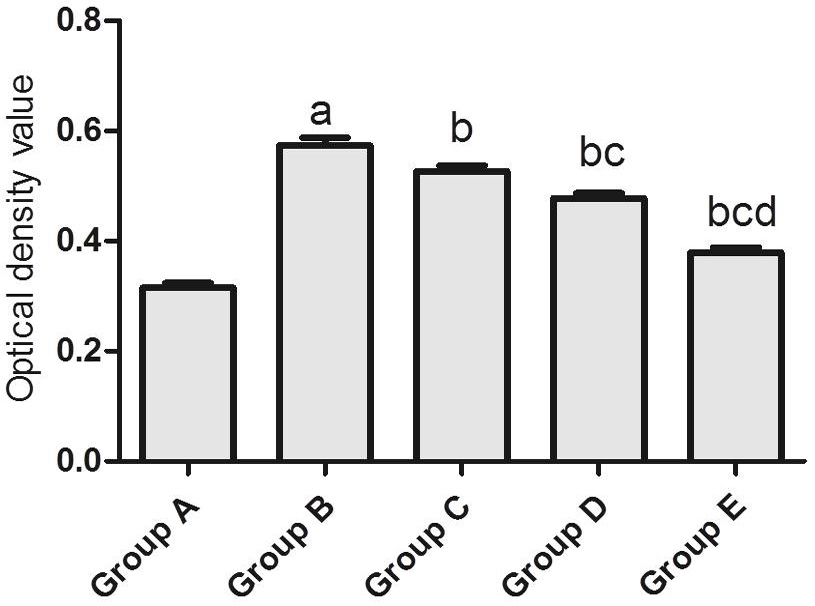
The effect of 5-FU on the OD value of bile duct scar fibroblasts in miniature pigs (n=3). Compared with group A, aP <0.05; compared with group B, bP <0.05; compared with group C, cP <0.05; and compared with group D, dP <0.05.
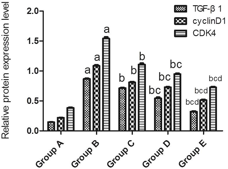
The effect of 5-FU on the expression levels of TGF-β1, cyclinD1, and CDK4 proteins in miniature pig bile duct scar fibroblasts (n=3).Compared with group A, aP <0.05; compared with group B, bP <0.05; compared with group C, cP <0.05; and compared with group D, dP <0.05.
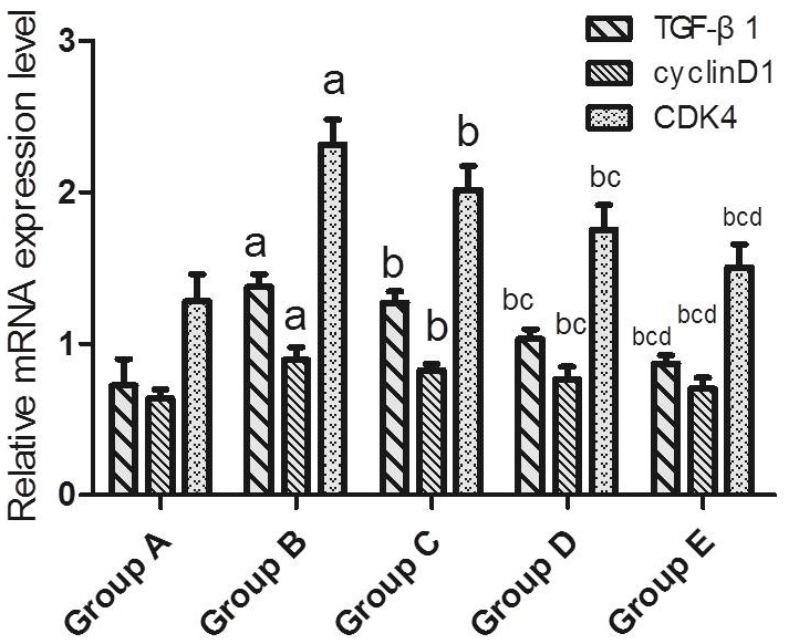
The effect of 5-FU on the expression levels of TGF-β1, cyclinD1, and CDK4 mRNA in miniature pig bile duct scar fibroblasts (n=3). Compared with group A, aP <0.05; compared with group B, bP <0.05; compared with group C, cP <0.05; and compared with group D, dP <0.05.
References
- Barsotti, C. and Ipata, P.L., 2002. Biochem. Pharmacol., 63: 117-122. https://doi.org/10.1016/S0006-2952(01)00845-0[CrossRef]
- Chi, C., Liu, X., Hou, F., Yu, X., Li, C., Cui, L., Liu, R. and Yin, C., 2018. BMC Complement. Altern. Med., 18: 52-60. https://doi.org/10.1186/s12906-018-2119-7[CrossRef]
- Fang, X., Hu, X., Zheng, Z., Tao, K., Wang, H., Guan, H., Shi, J., Ji, P., Cai, W., Bai, X., Zhu, X., Han, J., Liu, J. and Hu, D., 2019. J. mol. Histol., 50: 503-514. https://doi.org/10.1007/s10735-019-09844-w[CrossRef]
- Geng, Z.M., Yao, Y.M., Liu, Q.G., Niu, X.J. and Liu, X.G., 2005. World J. Gastroenterol., 11: 293-295. https://doi.org/10.3748/wjg.v11.i2.293[CrossRef]
- Gong, Y.F., Zhang, X.M., Liu, F., Wang, Z.Z., Deng, X.F., Jiao, Y., Li, X.J. and Huang, X.Y., 2016. Eur. J. Pharmacol., 791: 647-654. https://doi.org/10.1016/j.ejphar.2016.09.034[CrossRef]
- Hietanen, K.E., Järvinen, T.A., Huhtala, H., Tolonen, T.T., Kuokkanen, H.O. and Kaartinen, I.S., 2019. J. Plast. Reconstr. Aesthet. Surg., 2: 4-11. https://doi.org/10.1016/j.bjps.2018.05.052[CrossRef]
- Ji, X., Tang, Z., Shuai, W., Zhang, Z., Li, J., Chen, L., Cao, J. and Yin, W., 2019. Life Sci., 231: 116674. https://doi.org/10.1016/j.lfs.2019.116674[CrossRef]
- Maeda, T., Ebata, T., Yokoyama, Y., Igami, T., Mizuno, T., Yamaguchi, J., Onoe, S., Watanabe, N. and Nagino, M., 2019. Surg. Case Rep., 5: 177. https://doi.org/10.1186/s40792-019-0745-z[CrossRef]
- Ophir, A., 1991. Exp. Eye Res., 53: 799-803. https://doi.org/10.1016/0014-4835(91)90116-V[CrossRef]
- Wang, L., Cao, Y., Guan, Z., Luo, G., Luo, L., Yang, X. and Chu, M., 2020. Int. Urol. Nephrol., 52: 97-106. https://doi.org/10.1007/s11255-019-02286-z[CrossRef]
- Xu, J., Geng, Z.M. and Ma, Q.Y., 2003. Hepatobil. Pancreat. Dis. Int., 2: 295-299.
Services
Article usage statistics
Loading…